Is the temporal bone part of the orbit

Dylan Hughes
Published Apr 05, 2026
Walls of the Orbit The orbital roof consists of two parts of two bones, the orbital plate frontal bone and the lesser wing of the sphenoid bone. The fossa of the lacrimal gland is in the orbital roof, and it lies anteriorly and laterally behind the zygomatic process of the frontal bone.
Which bone is part of the orbit?
Walls of the Orbit The orbital roof consists of two parts of two bones, the orbital plate frontal bone and the lesser wing of the sphenoid bone. The fossa of the lacrimal gland is in the orbital roof, and it lies anteriorly and laterally behind the zygomatic process of the frontal bone.
Which bone does not form part of the orbit?
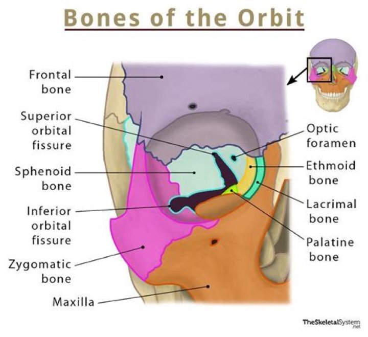
Which of the following bones does NOT form a part of the orbits of the eyes? Seven bones contribute to the orbits. They are the frontal, sphenoid, zygomatic, maxilla, palatine, lacrimal, and ethmoid bones. The vomer is not a contributor to the orbits.
What part of the skull is the orbit?
In anatomy, the orbit is the cavity or socket of the skull in which the eye and its appendages are situated. “Orbit” can refer to the bony socket, or it can also be used to imply the contents.Is the orbit part of the cranium?
The orbits are bony cavities on either side of the midsagittal plane of the skull below the cranium. They contain the globes, the extraocular muscles, and orbital nerves, blood vessels, and connective tissue.
What is the right orbit?
This image of the right orbit shows the 7 bones that contribute to its structure. The orbital process of the frontal bone and the lesser wing of the sphenoid form the orbital roof. The orbital plate of the maxilla joins the orbital plate of the zygoma and the orbital plate of the palatine bones to form the floor.
What bone contains large sinus inferior to orbit?
Structure. The ethmoid bone is an anterior cranial bone located between the eyes. It contributes to the medial wall of the orbit, the nasal cavity, and the nasal septum.
Where is the orbital bone around the eye?
The inferior wall, or orbital floor, is formed by the upper jawbone (maxilla), part of the cheek bone (zygomatic), and a small part of the hard palate (palatine bone). Fractures to the inferior floor most commonly come from a blow to the side of the face.What are orbital tissues?
The periorbita, also called the orbital periosteum or orbital fascia, covers the bones of the orbit (Figure 8-17). This dense connective tissue membrane serves as an attachment site for muscles, tendons, and ligaments and is a support structure for the blood supply to the orbital bones.
Where is the left orbit?The orbital rim is formed superiorly by the frontal bone, laterally by the zygomatic bone, inferiorly by the maxilla, and medially by portions of the frontal and maxillary bones.
Article first time published onWhat connects occipital and temporal bones?
Occipitomastoid suture. Connects occipital and temporal bones.
Which bone is not part of the eye orbit quizlet?
A small portion of the palatine bone articulates with the lateral mass of the ethmoid and is included in the orbital complex. The nasal bone is not included in the orbit.
Which of the following is not part of the axial skeleton?
The E) pelvic girdle is not part of the axial skeleton. This is part of the lower limbs, according to common practice.
How do you remember the bones of orbit?
- My Little Eye Sits (in the orbit); or.
- Medial Layer Eye Socket.
What are orbits in the brain?
What is an Orbit? An orbit refers to the boney cavity occupied by your eye, nerves, muscles, fat, and additional soft tissues needed for proper eye movement and function. They are symmetrical and separated by the nasal cavity and paranasal sinuses. An orbit MRI scan is sometimes also referred to as an orbital MRI scan.
Is the inferior nasal concha part of the ethmoid bone?
The inferior nasal conchae are a pair of bones, with one concha on either side, that separates the middle and lower nasal meatus, or nasal cavity. … While the superior and middle nasal conchae are technically part of the ethmoid bone, the inferior nasal concha forms a completely separate bone.
Does the temporal bone connects to the zygomatic bone via the temporal process of the temporal bone?
The temporal bone connects to the zygomatic bone via the temporal process of the temporal bone. … The frontal bone articulates with the parietal bone by means of the sagittal suture.
What bones form forehead?
The frontal bone forms the forehead. The two parietal bones form the upper sides of the skull; the two temporal bones form the lower sides. The butterfly-shaped sphenoid bone is located at the base of the skull.
Where is the orbital bone on your face?
Also called “the orbit,” the orbital “bone” is actually seven strong bones that make up the encasing of the open socket of the eye; these bones come together to house the actual eye. The periorbital skin is the skin/area around your eye.
What are the bones around the eye called?
There are seven orbital bones that make up this structure: the frontal, sphenoid, zygomatic, ethmoid, lacrimal, palatine and maxilla bones. Each of these plays a role in keeping the eyeball protected.
Where is optic foramen?
The optic foramen, the opening through which the optic nerve runs back into the brain and the large ophthalmic artery enters the orbit, is at the nasal side of the apex; the superior orbital fissure is a larger hole through which pass large veins and nerves.…
How do you fix a broken orbital bone?
- Remove bone fragments.
- Free trapped eye muscles and eliminate double vision.
- Restore the normal architecture of the eye socket if your injured eye looks sunken in.
- Repair deformities of the eye rim that affect your appearance.
How deep is an eye socket?
This distance varied from 4.4 to 5.7 cm in males (mean 5.024, SD 0.272) and from 4.5 to 5.5 cm in females (mean 4.9, SD 0.204).
Where is the orbital floor located?
The orbital floor, which forms the roof of the maxillary sinus, slopes upward toward the apex of the pyramid, which lies roughly 44 to 50 mm posterior to the orbital entrance [3,4]. This complicated anatomy makes repair and reconstruction of orbital fracture difficult for a novice (Fig. 1).
What muscles are attached to the temporal bone?
Muscular attachments The temporalis muscle originates from the temporal fossa, which is formed partially by the lateral aspect of the temporal bone. The sternocleidomastoid, splenius capitis, longissimus capitis and digastric are all attached to the mastoid process of the temporal bone.
What type of bone is the temporal bone?
The temporal bone is a thick, hard bone that forms part of the side and base of the skull. This bone protects nerves and structures in the ear that control hearing and balance.
Which bone is superior to the temporal bone?
Lateral to the arcuate eminences is the tegmen, a thin plate of bone roofing the mastoid antrum, epitympanic area, and external acoustic meatus. The temporal bone articulates anteriorly with the sphenoid bone, above with the parietal bone, and posteriorly with the occipital bone.
Which bone of the skull does not contain sinuses quizlet?
The nasal bones are small tombstone shaped bones that form the bridge of the nose and don’t contain sinuses. Identify the location of the sphenoid bone. The sphenoid bone spans the width of the floor of the skull.
Which of the following bones is not part of the lower limb?
42. Which of the following bones is NOT included within the lower limb? zygomatic and temporal bones.
Which bones do not contain a sinus?
Explanation: There are four paranasal sinuses in the head: the frontal, maxillary, sphenoid, and ethmoid sinuses. They function in lightening the skull, and creating mucous for the nasal cavity. The temporal bone does not contain a sinus.
Is the ilium part of the axial skeleton?
The clavicle, scapula, and bones of the pelvis (such as the ilium and pubis) are bones of the appendicular skeleton. The (e) Sacrum on the other hand forms part of the axial skeleton.