Is the ventral root part of the CNS

Andrew White
Published May 20, 2026
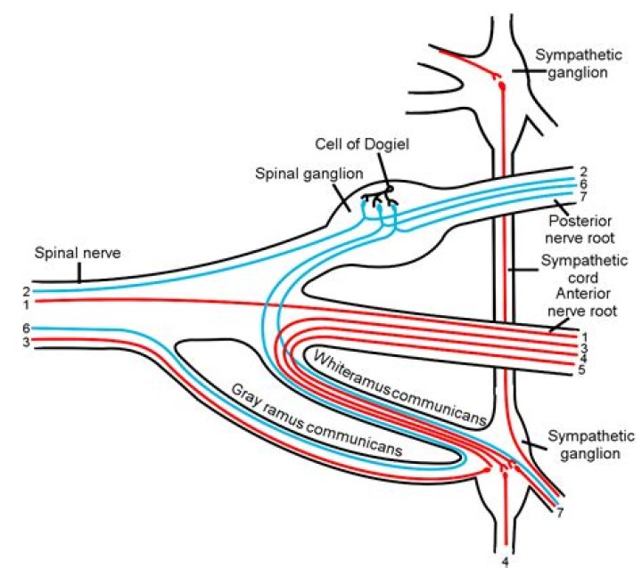
Ventral root of spinal nerveFMA5979Anatomical terminology
Is the ventral root in the CNS?
According to the available evidence, the presence of unmyelinated fibers in the ventral roots is explained as follows: afferent fibers from the periphery project into the central nervous system using ventral roots as a pathway to the spinal cord; afferent ventral root fibers innervate the ventral spinal pia mater and/ …
Is the dorsal root part of the CNS or PNS?
Dorsal nerve roots carry sensory neural signals to the central nervous system (CNS) from the peripheral nervous system (PNS). The dorsal root ganglion (DRG) has a significant clinical application, particularly in its association with neuropathic pain.
Are dorsal and ventral roots in the CNS?
Spinal Nerve. Each spinal nerve is connected to the spinal cord by the dorsal (sensory) and ventral (motor) nerve roots (Fig. 2.11). The axons of the basal plate neurons (motor neurons) pass through the marginal layer and form the ventral or motor root of spinal nerve.Are nerve roots part of CNS?
The nerve roots exit the spinal canal through the intervertebral foramen, small hollows between each vertebra. The brain and the spinal cord make up the Central Nervous System (CNS). The nerve roots that exit the spinal cord/spinal canal branch out into the body to form the Peripheral Nervous System (PNS).
What are ventral roots?
Medical Definition of ventral root : the one of the two roots of a spinal nerve that passes anteriorly from the spinal cord separating the anterior and lateral funiculi and that consists of motor fibers. — called also anterior root. — compare dorsal root.
Where is the ventral root?
the motor root of a spinal nerve, which carries motor information from the spinal cord to the rest of the body and leaves from the anterior side of the cord.
What are dorsal roots?
Definition of dorsal root : the one of the two roots of a spinal nerve that passes dorsally to the spinal cord and consists of sensory fibers.What is the dorsal root and ventral root?
Each spinal nerve has two roots, a dorsal or posterior (meaning “toward the back”) one and a ventral or anterior (meaning “toward the front”) one. The dorsal root is sensory and the ventral root motor; the first cervical nerve may lack the dorsal root. Oval swellings, the spinal ganglia, characterize the dorsal roots.
What does the ventral root of the spinal cord contain?The Ventral Root of the spinal nerve contains outgoing, efferent (meaning to “bear away from”) fibers that carry information destined to control motor or glandular function. The cell bodies of these motor neurons are located in the ventral horns of the spinal cord’s central grey region.
Article first time published onIs the ventral root afferent or efferent?
In anatomy and neurology, the ventral root, motor root or anterior root is the efferent motor root of a spinal nerve.
Does the ventral root contain sensory or motor neurons?
Ventral root fibers are the axons of motor and visceral efferent fibers and emerge from poorly defined ventral lateral sulcus as ventral rootlets. The ventral rootlets from discrete spinal cord section unite and form the ventral root, which contain motor nerve axons from motor and visceral motor neurons.
Where is the dorsal root?
the sensory root of a spinal nerve, which carries sensory information to the spinal cord and enters the posterior side of the cord. Spinal cord with arrow indicating a dorsal root.
What consists of the CNS?
The central nervous system is made up of the brain and spinal cord. The peripheral nervous system is made up of nerves that branch off from the spinal cord and extend to all parts of the body.
Are cranial nerves part of the CNS or PNS?
The cranial nerves are considered components of the peripheral nervous system (PNS), although on a structural level the olfactory, optic and terminal nerves are more accurately considered part of the central nervous system (CNS).
What is a sacral nerve root?
The sacral plexus (plexus sacralis) is a nerve plexus that provides motor and sensory nerves for the posterior thigh, most of the lower leg, the entire foot, and part of the pelvis (see the following image). … The sacral plexus is derived from the anterior rami of spinal nerves L4, L5, S1, S2, S3, and S4.
Is ventral horn sensory or motor?
The ventral horns contains the cell bodies of motor neurons that send axons via the ventral roots of the spinal nerves to terminate on striated muscles.
Is there ventral root ganglion?
why there is no ventral root ganglion .
What exits from the ventral root?
ventral roots (anterior roots) allow motor neurons to exit the spinal cord.
What is the ventral spinal cord?
Ventral roots consist of efferent fibers that arise from motor neurons whose cell bodies are found in the ventral (or anterior) gray horns of the spinal cord. The spinal cord (and brain) are protected by three layers of tissue or membranes called meninges, that surround the canal.
What are the function of ventral and dorsal roots in spinal cord?
Each spinal nerve is formed by the combination of nerve fibers from the dorsal and ventral roots of the spinal cord. The dorsal roots carry afferent sensory axons, while the ventral roots carry efferent motor axons.
Which structure makes the dorsal root different from the ventral root?
What structure makes the dorsal root different from the ventral root? The dorsal root has a ganglion. Muscles and glands that are capable of producing a response when stimulated by motor neurons are called …
What is the dorsal root of the spinal cord?
The dorsal root of spinal nerve (or posterior root of spinal nerve or sensory root) is one of two “roots” which emerge from the spinal cord. It emerges directly from the spinal cord, and travels to the dorsal root ganglion. Nerve fibres with the ventral root then combine to form a spinal nerve.
Where do the dorsal roots of the spinal nerves enter the spinal cord?
Dorsal and ventral roots enter and leave the vertebral column respectively through intervertebral foramen at the vertebral segments corresponding to the spinal segment. Drawing of the 8, 12, 5, 5 and 1 cervical, thoracic, lumbar, sacral and coccygeal spinal nerves and their exit from the vertebrate, respectively.
What type of neurons are found only in the ventral root of the spinal cord?
The anterior (ventral) root contains axons of motor neurons that conduct nerve impulses from the CNS to other parts of the body such as the muscles.
Are interneurons part of the CNS or PNS?
Interneurons (also known as association neurons) are neurons that are found exclusively in the central nervous system. ie Found in the brain and spinal cord and not in the peripheral segments of the nervous system.
What do the ventral roots and dorsal roots fuse to form what do the ventral roots and dorsal roots fuse to form?
These roots then leave the vertebral canal to lie within the intervertebral foramen. There, the dorsal and ventral roots fuse to form the spinal nerve, which is a mixed nerve carrying both sensory and motor fibers.
Are sensory signals are carried up the spinal cord or down the spinal cord?
Spinal Nerves The fibers of the sensory root carry sensory impulses to the spinal cord —pain, temperature, touch and position sense (proprioception)—from tendons, joints and body surfaces. The motor roots carry impulses from the spinal cord to the muscles.
Which neuron in a sensory pathway is part of the sensory receptor?
Structure. A somatosensory pathway will typically consist of three neurons: primary, secondary, and tertiary. In the periphery, the primary neuron is the sensory receptor that detects sensory stimuli like touch or temperature.
Where is the ventral horn located?
The ventral horns are at their largest in the area of the cervical and lumbar regions where they control muscle movement in the arms and legs.
Where is dorsal roots and ganglia located?
As the name indicates, the dorsal root ganglion is associated with the posterior or dorsal root of the spinal nerve. It is located in close proximity to the spinal cord. As the dorsal root of spinal nerve emerges from the intervertebral neural foramen, it expands to form the ganglion.