What cell bodies are located in the anterior horn

Rachel Hunter
Published May 23, 2026
one of the divisions of the grey matter of the spinal cord, the anterior horn contains cell bodies of alpha motor neurons
What does the anterior horns of the spinal cord contain?
The anterior horn of the spinal cord (also known as the anterior cornu) contains the cell bodies of motor neurons that affect the skeletal muscles.
What are anterior horns?
The term anterior horn (also frontal horn, anterior cornu, frontal cornu) may refer to either of two separate anatomical structures within the central nervous system: … anterior horn of spinal cord, the ventral (front) grey matter section of the spinal cord which contains motor neurons that affect the skeletal muscles.
What cell bodies are located in the lateral horn?
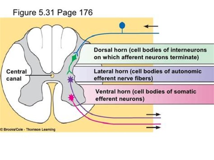
The lateral horn of the spinal cord is the small lateral projection of grey matter located between the dorsal horn and ventral horn and contain the neuronal cell bodies of the sympathetic nervous system.What types of cell bodies are found in the dorsal lateral and ventral horns?
The gray matter mainly contains the cell bodies of neurons and glia and is divided into four main columns: dorsal horn, intermediate column, lateral horn and ventral horn column.
Does the dorsal horn have cell bodies?
At the back of spinal cord the central grey matter forms two arms, each called a Dorsal Horn. The dorsal horns contain the cell bodies of sensory neurons. Two arms located at the front of the spinal cord, central grey matter are called ventral horns. They contain the cell bodies of motor neurons.
What are the anterior and posterior horns?
The posterior horn is responsible for sensory processing. The anterior horn sends out motor signals to the skeletal muscles. The lateral horn, which is only found in the thoracic, upper lumbar, and sacral regions, is the central component of the sympathetic division of the autonomic nervous system.
Are parasympathetic cell bodies in lateral horn?
The parasympathetic division of the ANS is sometimes called the craniosacral outflow because the cell bodies of presynaptic parasympathetic neurons are located in the brainstem (nuclei of cranial nerves III, VII, IX, and X) and in the lateral horn of spinal cord segments S2, S3, and S4.Where is the anterior gray horn?
Anterior horn cells (α-motor neurons), located in the anterior gray matter of the spinal cord, are found at every segment and are concentrated in the cervical and lumbosacral enlargements. Morphologic differentiation of the anterior horn cells is most evident from 12 to 14 weeks’ gestation.
Where is the ventral horn located?The ventral horns are at their largest in the area of the cervical and lumbar regions where they control muscle movement in the arms and legs.
Article first time published onIs the anterior horn part of the CNS or PNS?
A motor unit consists of an anterior horn cell, its motor axon, the muscle fibers it innervates, and the connection between them (neuromuscular junction). The anterior horn cells are located in the gray matter of the spinal cord and thus are technically part of the CNS.
What is ventral horn cells?
The ventral horn of the spinal cord is one of the grey longitudinal columns found within the spinal cord. It contains the cell bodies of the lower motor neurons which have axons leaving via the ventral spinal roots on their way to innervate muscle fibers.
Is the anterior horn the ventral horn?
The anterior grey column (also called the anterior cornu, anterior horn of spinal cord, motor horn or ventral horn) is the front column of grey matter in the spinal cord. It is one of the three grey columns. … The anterior grey column is the column where the cell bodies of alpha motor neurons are located.
Which neuron cell bodies are found in the anterior gray horns of the spinal cord?
one of the divisions of the grey matter of the spinal cord, the anterior horn contains cell bodies of alpha motor neurons, which innervate skeletal muscle to cause movement.
What is found in the anterior ventral root?
The ventral roots predominantly consist of efferent somatic motor fibers (thick alpha motor axons and medium-sized gamma motor axons) derived from nerve cells of the ventral column.
What specifically is found in the anterior gray horn of the spinal cord?
The ventral horn (also known as the anterior horn) largely contains motor neurons that exit the spinal cord to innervate skeletal muscle. The intermediate column and lateral horn contains neurons that innervate visceral and pelvic organs.
What is a posterior horn cell?
Neurons in the posterior (dorsal) horn of the spinal cord whose cell bodies and processes are confined entirely to the central nervous system. … They receive collateral or direct terminations of dorsal root fibers.
What are horns in spinal cord?
The ventral horns contains the cell bodies of motor neurons that send axons via the ventral roots of the spinal nerves to terminate on striated muscles.
Where is the posterior horn located?
The posterior horn of the medial meniscus is the posterior third of the medial meniscus. It is located in the back of the knee. It is the thickest portion and absorbs the most force, so therefore it provides the most stability to the knee and is the most important portion of the medial meniscus.
What neurons are in the dorsal horn?
…the spinal cord: (1) the dorsal horns, composed of sensory neurons, (2) the lateral horns, well defined in thoracic segments and composed of visceral neurons, and (3) the ventral horns, composed of motor neurons.
What are dorsal horn cells?
The dorsal horn functions as an intermediary processing center for this information, comprising a complex network of excitatory and inhibitory interneurons as well as projection neurons that transmit the processed somatosensory information from the spinal cord to the brain.
Which portion of the spinal cord contains cell bodies and axons?
The grey column, (as three regions of grey columns) in the center of the cord, is shaped like a butterfly and consists of cell bodies of interneurons, motor neurons, neuroglia cells and unmyelinated axons.
What type of cell bodies are found in the lateral gray horn?
The lateral horn of the spinal cord is the small lateral projection of grey matter located between the dorsal horn and ventral horn and contain the neuronal cell bodies of the sympathetic nervous system.
What type of cell bodies sensory motor Both are found in the posterior gray horn?
Posterior gray horn: Contains the cell bodies of visceral and somatic sensory neurons.
Are anterior horn cells lower motor neurons?
MOTOR NEURON DISEASE. The clinical hallmarks of anterior horn cell disease are the lower motor neuron signs of weakness, wasting (atrophy), and fasciculations.
Where are cell bodies of preganglionic sympathetic neurons located?
Anatomically, the sympathetic preganglionic neurons, the cell bodies of which are located within the central nervous system, originate in the lateral horns of the 12 thoracic and the first 2 or 3 lumbar segments of the spinal cord.
Where are the cell bodies of sympathetic preganglionic fibers located in the spinal cord?
The cell bodies of sympathetic preganglionic neurons are located in intermediolateral nuclei of the spinal cord. (A nucleus is a profile of a neuron column.) Each intermediolateral column forms a lateral gray horn in a thoracolumbar spinal cord transection.
Where would you find the cell bodies of preganglionic parasympathetic neurons?
Parasympathetic preganglionic neurons have cell bodies located in the brainstem and in the sacral (toward the bottom) spinal cord, as shown in Figure 16.27. The axons of the preganglionic neurons release acetylcholine on the postganglionic neurons, which are generally located very near the target organs.
Is ventral the same as anterior?
Directional Terms Anterior or ventral – front (example, the kneecap is located on the anterior side of the leg). Posterior or dorsal – back (example, the shoulder blades are located on the posterior side of the body).
Where is the anterior median fissure located?
The anterior median fissure (ventral or ventromedian fissure) contains a fold of pia mater, and extends along the entire length of the medulla oblongata: It ends at the lower border of the pons in a small triangular expansion, termed the foramen cecum.
Is the posterior horn part of the PNS?
The sacral spinal cord is at the level of the upper lumbar vertebral bones. The spinal nerves extend from their various levels to the proper level of the vertebral column.