What does the Thoracodorsal artery supply

Nathan Sanders
Published May 12, 2026
The thoracodorsal artery supplies predominantly the latissimus dorsi muscle
What is thoracodorsal artery?
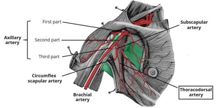
The thoracodorsal artery is one of the two terminal branches of the subscapular artery. It runs inferomedially along the lateral border of the scapula. Along its course, this artery gives rise to small branches that supply the following muscles: … The teres major muscle. The latissimus dorsi muscle and the adjacent skin.
What nerve runs with the thoracodorsal artery?
Thoracodorsal nerveLatissimus dorsiDetailsFromposterior cord (C6-C8)InnervatesLatissimus dorsi muscle
What does the thoracodorsal nerve innervate?
The thoracodorsal nerve is a pure motor nerve that innervates the latissimus dorsi muscle.What does circumflex scapular artery supply?
The circumflex scapular artery is the largest terminal branch of the subscapular artery. … They contribute to the blood supply of three muscles (deltoid, teres minor, triceps brachii), glenohumeral joint and two small cutaneous areas partially overlying the scapula.
What is thoracodorsal artery perforator flap?
The thoracodorsal artery perforator or TAP flap is a fasciocutaneous flap based on a musculocutaneous perforator or perforators from the thoracodorsal vessel axis and/or its vertical branch derivative. … A flap of dimensions 15 X 8 cm can be harvested on a single perforator.
Where does the thoracodorsal artery supply?
The thoracodorsal artery supplies predominantly the latissimus dorsi muscle but also gives branches to the serratus anterior muscle, the axillary skin, the subscapular and teres major muscles.
What does the ulnar nerve innervate?
The ulnar nerve innervates the flexor muscles of the forearm including the flexor carpi ulnaris and flexor digitorum profundus. It also innervates the intrinsic muscles of the hand including the palmaris brevis, lumbricals, hypothenar and interossei muscles.What happens with a Thoracodorsal nerve injury?
An intact thoracodorsal nerve may also cause atrophy of the muscle, which can lead to shoulder and arm weakness that hampers numerous common movements, including standing up from a chair.
Which muscles arises from the Thoracodorsal fascia and forms the posterior axillary fold?The teres major muscle lies superior to latissimus dorsi. The adjoining fibers of these two muscles are united, but separated by a bursa towards their humeral attachments. As they span the interval between the scapula and proximal humerus, the latissimus dorsi and teres major form the posterior axillary fold.
Article first time published onWhat does lateral thoracic artery supply?
The lateral thoracic artery supplies the axillary lymph nodes, serratus anterior, pectoralis major, pectoralis minor and subscapularis muscles. … Additionally, it can provide blood supply to the breast in females.
What are the alternate names of the thoracodorsal nerve?
The thoracodorsal nerve also known as the middle subscapular or long subscapular nerve arises from the posterior cord of the brachial plexus and supplies the latissimus dorsi muscle.
What is scapular anastomosis?
The scapular anastomosis is a system connecting certain subclavian artery and their corresponding axillary artery, forming a circulatory anastomosis around the scapula. It allows blood to flow past the joint in case of occlusion, damage, or pinching of the following scapular arteries: Transverse cervical artery.
What does the circumflex humeral artery supply?
Supply. The anterior circumflex humeral artery provides part of the blood supply to the glenohumeral joint, teres major and minor, and deltoid muscles. The ascending branch provides supply to the head of the humerus 1.
What does the Suprascapular artery supply?
The suprascapular artery supplies the supra- and infraspinatus muscles and travels with the suprascapular nerve. It is a direct branch from the subclavian artery (12%), the thyrocervical trunk (27%) or the internal thoracic artery (11%).
What does circumflex scapular artery Anastomose with?
It enters the infraspinatous fossa under cover of the Teres minor, and anastomoses with the transverse scapular artery (suprascapular) and the descending branch of the transverse cervical (a.k.a. dorsal scapular artery).
Where is the radial artery?
The radial artery runs on the inside of the forearm from the elbow to the thumb. The artery lies just under the surface of the skin. You may be able to see the blue or purple vein inside your wrist where the artery brings blood to the thumb.
What is the ulnar artery?
The ulnar artery is the last branch, or the terminal branch, of the brachial artery. It transports oxygenated blood to each of the muscles in the forearm and the hand.
What is the occipital artery a branch of?
The occipital artery arises in the neck anterior to the mastoid protuberance from the posterior aspect of the carotid. It branches from the carotid just a little above the facial artery (Fig. 17.1).
What is an artery perforator?
A perforator flap is a flap consisting of skin and subcutaneous tissue that is perfused by an isolated artery and vein that perforates the deep tissues (muscle or fascia).
What is Licap surgery?
Lateral intercostal artery perforator flap breast reconstruction (LICAP) aims to restore the size and shape of your breast by replacing lost breast tissue from the cancer surgery with skin and fat taken from the side of your chest wall and back.
What is Tdap flap?
Thoracodorsal Artery Perforator (TDAP) Flap The TDAP flap is a modified flap based on the traditional latissimus dorsi myocutaneous flap. It is raised as a fasciocutaneous flap based on a medial or lateral branch of the thoracodorsal arteries, drained by two venae comitantes. The blood supply is constant and reliable.
What is the function of the teres major?
The main function of teres major is to produce the movements of the humerus at the glenohumeral joint; it pulls the anterior surface of the humerus medially towards the trunk (internal rotation). Furthermore, it can extend the arm from the flexed position.
What Innervates rhomboid major?
The rhomboids are important in upper limb movement and stability of both the shoulder girdle and scapula. Both rhomboids receive innervation from the dorsal scapular nerve and supplied by the dorsal scapular artery.
What does the lower Subscapular nerve innervate?
The lower subscapular nerve (C5-C6) innervates teres major; and both the upper and lower subscapular nerves innervate the subscapularis, the third muscle of the rotator cuff apparatus. The thoracodorsal nerve (C6-C8), which also arises from the posterior cord, innervates the levator scapulae.
What does the superficial branch of the ulnar nerve supply?
The superficial branch of the ulnar nerve supplies sensory innervation to the anterior aspect of the ulnar 1½ digits (little finger and half of the ring finger) and medial palmar skin. Additionally, it provides motor innervation to the palmaris brevis muscle in the hypothenar region of the hand.
What does the axillary nerve innervate?
The axillary nerve supplies three muscles in the arm: deltoid (a muscle of the shoulder), triceps (long head) and teres minor (one of the rotator cuff muscles).
What does median nerve supply?
The median nerve innervates many muscles of the anterior forearm and hand, providing signals to and from the brain and spinal cord. The flexor digitorum superficialis and pronator quadratus are among the muscles of the anterior forearm that are solely innervated by the median nerve.
What are spinal erectors?
The erector spinae muscles are a group of long muscles that originate near the sacrum and extend vertically up the length of the back. The erector spinae muscles lie on each side of the vertebral column and extend alongside the lumbar, thoracic, and cervical sections of the spine.
Which muscles make up the posterior abdominal wall?
The posterior abdominal wall primarily serves as protection for the retroperitoneal organs. It is mostly muscular contributed by the diaphragm, paraspinal, quadratus lumborum, iliacus, and psoas muscles.
What are the posterior abdominal muscle?
The posterior abdominal muscles are quadratus lumborum, psoas major, psoas minor, and iliacus. Knowledge of these muscles, especially the pass major, is vital to the lateral transpsoas spine surgeon. This chapter discusses the normal and variant anatomy of these muscles.