What is a clinical manifestation of hypernatremia in Burns

Rachel Hunter
Published Feb 26, 2026
Symptoms of hypernatremia tend to be nonspecific. Anorexia, restlessness, nausea, and vomiting occur early. These symptoms are followed by altered mental status, lethargy or irritability, and, eventually, stupor or coma.
What is a clinical manifestation of hypernatremia in Burns? - Google Search
Nonspecific symptoms like lightheadedness, nausea, headache, fatigue, and confusion may be consistent in alert hypernatremic patients4; in unconscious patients, laboratory results have to be observed vigilantly because of the lack of these symptoms.
Which feature is associated with maturation phase of wound healing?
During the maturation phase, collagen is aligned along tension lines and water is reabsorbed so the collagen fibers can lie closer together and cross-link. Cross-linking of collagen reduces scar thickness and also makes the skin area of the wound stronger.
What complications may be caused by sepsis in Burns?
- a high temperature.
- dizziness.
- vomiting.
Which feature is associated with the maturation phase?
In the maturation phase, the dominant feature is collagen. The dense bundle of fibers, characteristic of collagen, is the predominant constituent of the scar. Wound contraction occurs to some degree in primary closed wounds but is a pronounced feature in wounds left to close by secondary intention.
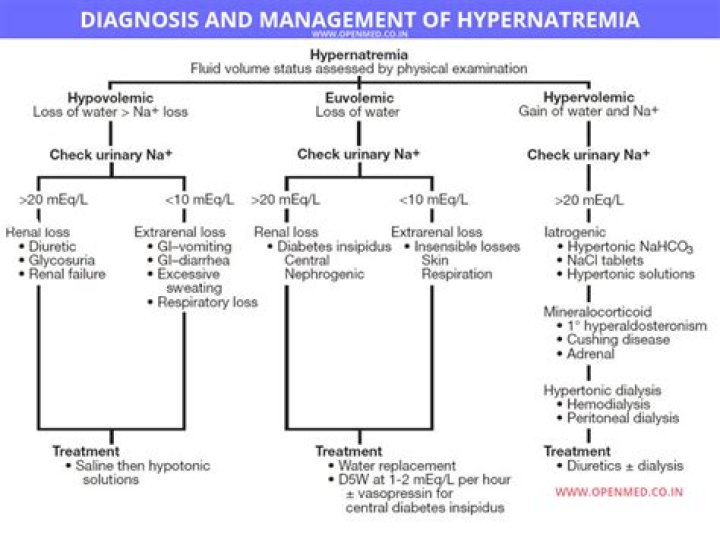
What is the most common cause of hypernatremia?
Two common causes of hypernatremia are insufficient fluid intake and too much water loss. In rare cases, consuming too much sodium can cause hypernatremia to occur. The opposite of hypernatremia is hyponatremia. This condition occurs when a person’s serum sodium level is less than 135 mEq/l.
Do burns cause hypernatremia?
In critically ill burn patients, hypernatremia is a common condition and can occur in up to 11% of severely burned patients.
What does it mean when a burn turns purple?
Tell-Tale Signs of Infected Burn As the skin around a burn blisters and fills with fluid, it becomes vulnerable to infection. If you see or experience the following, you could have an infection: Any change in color of the burnt area or the skin surrounding it. Swelling with purplish discoloration.What are some common burn complications?
- Bacterial infection, which may lead to a bloodstream infection (sepsis)
- Fluid loss, including low blood volume (hypovolemia)
- Dangerously low body temperature (hypothermia)
- Breathing problems from the intake of hot air or smoke.
- Scars or ridged areas caused by an overgrowth of scar tissue (keloids)
drench the burn thoroughly with cool water to prevent further damage and remove all burned clothing. If the burn area is limited, immerse the site in cold water for 30 minutes to reduce pain and oedema and to minimize tissue damage.
Article first time published onWhat are signs of wound healing?
- The wound becomes slightly swollen, red or pink, and tender.
- You also may see some clear fluid oozing from the wound. …
- Blood vessels open in the area, so blood can bring oxygen and nutrients to the wound. …
- White blood cells help fight infection from germs and begin to repair the wound.
What are the 3 classifications of wound healing?
Primary healing, delayed primary healing, and healing by secondary intention are the 3 main categories of wound healing.
What is the difference between regeneration and fibrosis?
Damage-induced matrix deposition is a transient phenomenon of the regenerative response, and successful healing entails its eventual removal (61, 62). Fibrosis occurs when ECM proteins accumulate in excessive amounts, leading to scarring that distorts the normal layout and stiffness of the tissue.
Which diagnostic test may be used to distinguish vascular from nonvascular structures?
Magnetic resonance imaging is used for distinguishing vascular from nonvascular structures. An X-ray is useful to screen, diagnose, and evaluate changes in the respiratory system.
What is remodeling in wound healing?
Remodeling or also known as maturation phase is the fourth and final phase in wound healing and lasts from 21 days up to 2 years. In this final and longest phase, collagen synthesis is ongoing in order to strengthen the tissue. Remodeling occurs as wound continues to contract and fibers are being reorganized.
Which skin color alteration may be observed in a client diagnosed with methemoglobinemia?
Symptoms are proportional to the methemoglobin level and include skin color changes (cyanosis with blue or grayish pigmentation) and blood color changes (brown or chocolate color). As levels of methemoglobin rise above 15%, neurologic and cardiac symptoms arise as a consequence of hypoxia.
What causes hyperkalemia in Burns?
Introduction: Classically, hyperkalemia has been regarded as a complication in patients with electrical burns. The etiology of hyperkalemia includes metabolic acidosis, destruction of red blood cells, rhabdomyolysis and the development of renal failure.
What happens to sodium in burn patients?
Abstract. Following burn injury, as after other forms of trauma, there is renal sodium and water retention with increased urinary potassium losses. The hyponatræmia in these cases results rarely from sodium deficit but usually from excess water retention and entry of sodium into the cells.
What electrolyte imbalance is caused by burns?
Hyponatraemia is frequent, and the restoration of sodium losses in the burn tissue is therefore essential hyperkalaemia is also characteristic of this period because of the massive tissue necrosis. Hyponatraemia (Na) (< 135 mEq/L) is due to extracellular sodium depletion following changes in cellular permeability.
How is hypernatremia diagnosed?
Diagnosis. Hypernatremia is often diagnosed through blood tests. Urine tests can also be used to identify high levels of sodium along with urine concentration. Both blood and urine tests are fast, minimally invasive tests that require no preparation.
How does heat stroke cause hypernatremia?
Hypernatremia can be caused by pure water loss and dehydration, while hyponatremia can be caused by increased hypertonic fluid loss (e.g., vomiting, diarrhea). Hyperkalemia can be seen as a result of cellular death, particularly with rhabdomyolysis.
What causes the clinical manifestations of severe symptomatic hypophosphatemia?
The major conditions associated with symptomatic hypophosphatemia are chronic alcoholism, intravenous hyperalimentation without phosphate supplementation, urinary phosphate-wasting syndromes (such as Fanconi syndrome or tumor-induced osteomalacia), and the chronic ingestion of antacids or other phosphate binders.
What is the pathophysiology of a burn?
The pathophysiology of the burn wound is characterized by an inflammatory reaction leading to rapid oedema formation, due to increased microvascular permeability, vasodilation and increased extravascular osmotic activity.
What are the effects of burns?
The effects of burns These may include loss of limb(s), disfigurement, loss of mobility, scarring, and recurrent infections. Severe burns can also penetrate deep skin layers. This can cause muscle or tissue damage that may affect every system of the body.
What are long term complications of burns?
Major burns may have long lasting impact on the quality of people’s lives, with persisting problems related to scarring, contractures, weakness, thermoregulation, itching, pain, sleep, body image and psychosocial wellbeing.
What does 3rd degree burn look like?
A third-degree burn will not produce blisters or look wet. Instead, it will look dark red, dry, and leathery. Touching a third-degree burn usually does not cause pain. You will easily be able to see that the burn penetrates deeply into the skin, and you may even see yellowish, fatty tissue in the wound bed.
What are the three classifications of burns?
- First-degree (superficial) burns. First-degree burns affect only the outer layer of skin, the epidermis. …
- Second-degree (partial thickness) burns. …
- Third-degree (full thickness) burns. …
- Fourth-degree burns.
How do you know when a burn is infected?
- an increase in pain or discomfort around the affected area.
- redness in the area of the burn, especially if it begins to spread or form a red streak.
- swelling or warmth in the affected area.
- fluid or pus oozing from the burn site.
- a bad smell around the burn.
What are the medical interventions to be expected for a burn patient?
For serious burns, after appropriate first aid and wound assessment, your treatment may involve medications, wound dressings, therapy and surgery. The goals of treatment are to control pain, remove dead tissue, prevent infection, reduce scarring risk and regain function.
What is the initial treatment for a burn?
Hold the burned area under cool (not cold) running water or apply a cool, wet compress until the pain eases. Remove rings or other tight items from the burned area. Try to do this quickly and gently, before the area swells. Don’t break blisters.
What are the 6 C's of Burn Care?
Burns are now commonly classified as superficial, superficial partial thickness, deep partial thickness and full thickness. A systematic approach to burn care focuses on the six “Cs”: clothing, cooling, cleaning, chemoprophylaxis, covering and comforting (i.e., pain relief).