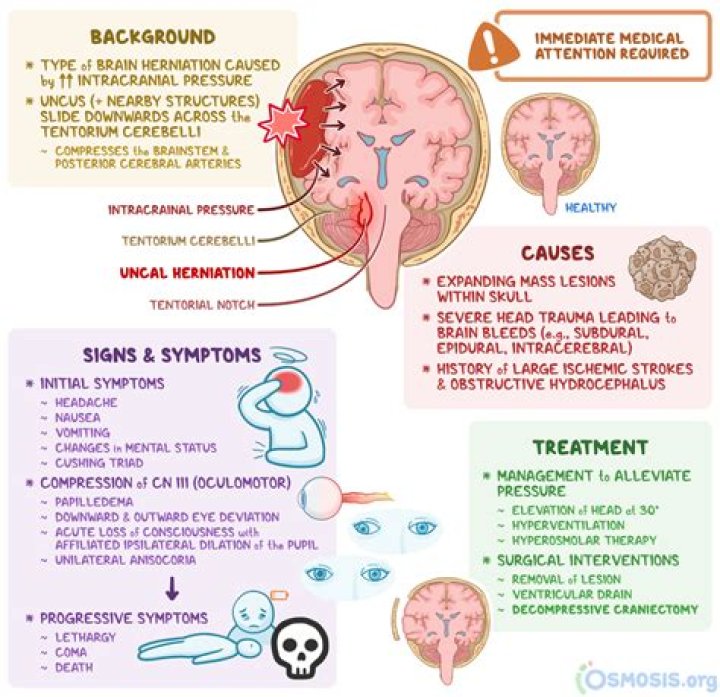
What is an uncal herniation

Rachel Hunter
Published Mar 28, 2026
Uncal herniation occurs when rising intracranial
Which brain herniation is the most life threatening?
Central herniation Downward herniation can stretch branches of the basilar artery (pontine arteries), causing them to tear and bleed, known as a Duret hemorrhage. The result is usually fatal.
What is the most common brain herniation?
Subfalcine hernia, also known as midline shift or cingulate hernia, is the most common type of cerebral hernia. It is generally caused by unilateral frontal, parietal, or temporal lobe disease that creates a mass effect with medial direction, pushing the ipsilateral cingulate gyrus down and under the falx cerebri.
Can a person survive brain herniation?
The outlook varies, depending on where in the brain the herniation occurs. Without treatment, death is likely. There can be damage to parts of the brain that control breathing and blood flow. This can rapidly lead to death or brain death.What are the types of brain herniation?
- Subfalcine herniation.
- Transalar (transsphenoidal) herniation.
- Transtentorial uncal herniation.
- Central (trans-tentorial) herniation (descending and ascending)
- Cerebellar tonsillar herniation.
- Transcalvarial herniation.
How does brain herniation cause death?
Pressure then builds up and causes a decrease in blood flow as well as damage to the tissues. This pressure and swelling causes death by a process called ‘coning’ where the brain is forced through a small opening at the base of the skull where it meets the spinal cord.
What causes uncal herniation?
Uncal herniation occurs when rising intracranial pressure causes portions of the brain to move from one intracranial compartment to another. It is a life-threatening neurological emergency and indicates the failure of all adaptive mechanisms for intracranial compliance.
What is cerebellar herniation?
A tonsillar herniation is characterized by the descent of the cerebellar tonsils through the foramen magnum, which compresses the medulla against the clivus/odontoid process. It is described as “coning” as the brain tissue is squeezed down through the foramen like being squeezed into a cone. (Where is the Uncus?
The uncus is located in the inferior, medial aspect of the temporal lobe. This structure, and the adjacent parahippocampal gyrus, can herniate through the tentorial notch. This is most commonly seen with smaller temporal lesions or larger lesions involving the frontal or parietal lobes.
What is kernohan notch?Kernohan’s notch phenomenon is the ipsilateral hemiplegia caused by compression of the contralateral cerebral peduncle against the tentorial edge by a supratentorial mass. Diffusion tensor imaging (DTI) and transcranial magnetic stimulation (TMS) could be useful for exploring the state of the corticospinal tract (CST).
Article first time published onWhat is the importance of Transtentorial herniation?
A transtentorial herniation is the movement of brain tissue from one intracranial compartment to another. This includes uncal, central, and upward herniation. These are life-threatening and time-critical pathologies that may be reversible with emergent surgical intervention and medical management.
When does Transtentorial herniation occur?
Upward transtentorial herniation can occur when an infratentorial mass (eg, tumor in the posterior fossa, cerebellar hemorrhage) compresses the brain stem, kinking it and causing patchy brain stem ischemia. The posterior 3rd ventricle becomes compressed.
Which of the following terms means herniation of the brain?
Craniocele: The herniation of brain substance through the skull. Cranioplasty: The surgical repair of the skull.
How does uncal herniation produce ipsilateral hemiparesis?
The mass lesion causing the uncal herniation usually causes a contralateral hemiparesis, but as the pressure increases, the opposite cerebral peduncle is compressed against the tentorium, which causes an ipsilateral hemiparesis (Kernohan’s sign).
What is ipsilateral pupil dilation?
A unilateral, ipsilateral (on the same side as the lesion), fixed dilated pupil is the initial focal sign, followed by bilateralfixed dilated pupils, occurring anything from minutes to hours later.
Can a person live with brain stem damage?
What happens when you damage your brain stem. When an accident causes brain stem damage, the affects can be devastating. In fact, destruction of the midbrain, pons, or medulla oblongata causes “brain death”, and the unfortunate victim of the injury cannot survive.
Can a brain stem heal?
The brain stem is home to the most basic life functions, and the resulting damage can be devastating. However, it is possible for a person with a brain stem injury to at least partially recover by using the brain’s natural plasticity.
Can you breathe on your own if you are brain dead?
It can be confusing to be told someone has brain death, because their life support machine will keep their heart beating and their chest will still rise and fall with every breath from the ventilator. But they will not ever regain consciousness or start breathing on their own again.
Is the Uncus a part of the olfactory cortex?
The part of the olfactory cortex that is on the temporal lobe covers the area of the uncus, which leads into the two significant clinical aspects of the uncus: uncinate fits and uncal herniations.
What does the hippocampus do?
Hippocampus is a complex brain structure embedded deep into temporal lobe. It has a major role in learning and memory. It is a plastic and vulnerable structure that gets damaged by a variety of stimuli. Studies have shown that it also gets affected in a variety of neurological and psychiatric disorders.
What does the piriform cortex do?
The piriform cortex serves a critical role in odor discrimination and perception, synthetic processing of complex odorant mixtures, experience- and state-dependent olfactory sensory gating, short-term odor habituation, and odor memory.
What is in the brain stem?
The brainstem is the structure that connects the cerebrum of the brain to the spinal cord and cerebellum. It is composed of 3 sections in descending order: the midbrain, pons, and medulla oblongata.
Why would you need a craniectomy?
A craniectomy is a surgery done to remove a part of your skull in order to relieve pressure in that area when your brain swells. A craniectomy is usually performed after a traumatic brain injury. It’s also done to treat conditions that cause your brain to swell or bleed.
What is true about crus cerebri?
The cerebral crus (crus cerebri) is the anterior portion of the cerebral peduncle which contains the motor tracts, travelling from the cerebral cortex to the pons and spine. The plural of which is cerebral crura.
What is Duret hemorrhage?
Duret haemorrhages are small linear areas of bleeding in the midbrain and upper pons of the brainstem. They are caused by a traumatic downward displacement of the brainstem. They are named after Henri Duret.
What does the cerebral peduncle do?
As a whole, the cerebral peduncles assist in refining motor movements, learning of new motor skills, and converting proprioceptive information into balance and posture maintenance. Important fiber tracts that run through the cerebral peduncles are the corticospinal, corticopontine, and corticobulbar tracts.
What is the Supratentorial?
The supratentorial area (the upper part of the brain) contains the cerebrum, lateral ventricle and third ventricle (with cerebrospinal fluid shown in blue), choroid plexus, pineal gland, hypothalamus, pituitary gland, and optic nerve. … The skull and meninges protect the brain and spinal cord (left panel).
What are cerebellar tonsils?
Anatomical terms of neuroanatomy. The cerebellar tonsil (Latin: tonsilla cerebelli) is analogous to a rounded lobule on the undersurface of each cerebellar hemisphere, continuous medially with the uvula of the cerebellar vermis and superiorly by the flocculonodular lobe.
What is radiography of the brain called?
Magnetic resonance imaging (MRI) of the brain is a safe and painless test that uses a magnetic field and radio waves to produce detailed images of the brain and the brain stem.
What causes brain stem swelling?
Fluid collection within the brain tissue, called cerebral edema, can result from numerous causes, including infections, trauma, stroke, brain tumors, certain toxic substances, complications of diabetes, chemical imbalances, abuse of opioids, extreme high blood pressure (malignant hypertension), or high altitude …
What should brain pressure?
For the purpose of this article, normal adult ICP is defined as 5 to 15 mm Hg (7.5–20 cm H2O). ICP values of 20 to 30 mm Hg represent mild intracranial hypertension; however, when a temporal mass lesion is present, herniation can occur with ICP values less than 20 mm Hg [5].