What is conjunctival hyperemia

Lily Fisher
Published Mar 01, 2026
Conjunctival hyperaemia is one of the most common findings in ophthalmologic practice. It is routinely described as a symptom of many ocular diseases such as conjunctivitis, uveitis, elevated intraocular pressure due to glaucoma, and ophthalmic side effects.
Is conjunctival hyperemia bad?
Although conjunctival hyperemia is an important clinical sign of ocular disease or inflammation, it is important to note that even a normal eye has a degree of hyperemia; it is more common in males than females; and the area of the nasal bulbar has the highest grading.
What is conjunctival irritation?
When the conjunctiva is inflamed, the white of the eye appears red. Conjunctivitis is the most likely diagnosis when a patient has a red eye and discharge. Irritant conjunctivitis is a non-infectious form of conjunctivitis caused by a transient mechanical or chemical insult. It can be acute, relapsing or chronic.
What is a conjunctival?
The conjunctiva is the clear, thin membrane that covers part of the front surface of the eye and the inner surface of the eyelids. It has two segments: Bulbar conjunctiva. This portion of the conjunctiva covers the anterior part of the sclera (the “white” of the eye).What are conjunctival diseases?
Pink eye (conjunctivitis) is an inflammation or infection of the transparent membrane (conjunctiva) that lines your eyelid and covers the white part of your eyeball. When small blood vessels in the conjunctiva become inflamed, they’re more visible. This is what causes the whites of your eyes to appear reddish or pink.
How do you Recognise hyperaemia?
- dark blue or red tinge.
- swollen.
- cooler than usual to touch.
- in chronic cases, brown in color.
How is conjunctival hyperemia treated?
Symptoms are conjunctival hyperemia and ocular discharge and, depending on the etiology, discomfort and itching. Diagnosis is clinical; sometimes cultures are indicated. Treatment depends on etiology and may include topical antibiotics, antihistamines, mast cell stabilizers, and corticosteroids.
What keeps conjunctiva moist?
Small glands at the edge of the upper and lower eyelids secrete an oily substance that contributes to the tear film and keeps tears from evaporating. Tears keep the surface of the eye moist. Without such moisture, the normally transparent cornea can become dried, injured, infected, and opaque.Where is the conjunctival sac of the eye?
The conjunctival sac is the space bound between the palpebral and bulbar conjunctiva in to which the lacrimal fluid is secreted and opens interiorly between the eyelids. it ends at the superior and inferior conjunctival fornices.
What are the whites of your eyes called?Sclera: the white of your eye. Conjunctiva: a thin layer of tissue that covers the entire front of your eye, except for the cornea.
Article first time published onWhy does it look like I have a bubble in my eye?
Chemosis is a sign of eye irritation. The outer surface of the eye (conjunctiva) may look like a big blister. It can also look like it has fluid in it. When severe, the tissue swells so much that you can’t close your eyes properly.
Does Covid start with red eyes?
Based on data so far, doctors believe that 1%-3% of people with COVID-19 will get conjunctivitis, also called pinkeye. It happens when the virus infects a tissue called conjunctiva, which covers the white part of your eye or the inside of your eyelids. Symptoms include if your eyes are: Red.
What is a ciliary flush?
Ciliary flush is usually present in eyes with corneal inflammation, iridocyclitis or acute glaucoma, though not simple conjunctivitis. A ciliary flush is a ring of red or violet spreading out from around the cornea of the eye.
When is conjunctivitis no longer contagious?
Pink eye (conjunctivitis) generally remains contagious as long as your child is experiencing tearing and matted eyes. Signs and symptoms of pink eye usually improve within three to seven days. Check with your doctor if you have any questions about when your child can return to school or child care.
What is the best antibiotic for eye infection?
Patients with symptoms should be referred immediately to an ophthalmologist. Oral antibiotics such as azithromycin or doxycycline are effective treatments.
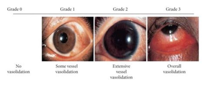
What does conjunctival hyperemia look like?
Conjunctival hyperemia is a conjunctival reaction that appears as dilation and redness of the conjunctival vessels. The pattern of hyperemia often appears with the greatest redness at the fornices and fades moving toward the limbus.
What is metabolic hyperaemia?
Functional hyperaemia, metabolic hyperaemia, arterial hyperaemia or active hyperaemia, is the increased blood flow that occurs when tissue is active. Hyperaemia is likely mediated by the increased synthesis and/or release of vasodilatory agents during periods of heightened cellular metabolism.
Why is my eye half red on the bottom?
Ocular drug-induced allergic reaction. A redness in the lower half of the eye is often a sign of allergic reaction to a topical ophthalmic agent.
Why is hyperemia an active process?
Hyperemia is an active engorgement of vascular beds with a normal or decreased outflow of blood. It occurs because of increased metabolic activity of tissue that results in localized increased concentrations of CO2, acid, and other metabolites.
Which of the following will trigger hyperemia?
Hyperemia is the increase of blood to your organs. There are two types of hyperemia. The causes of hyperemia include exercise, digestion, fever, hot flashes, injury and infection, heart failure, and thrombosis. Hyperemia is the increase of blood to your organs.
What's the difference between hyperaemia and erythema?
Erythemia is redness of skin or mucous membrane caused by Hyperemia, which is the presence of increased blood flow to a particular structure. Erythema is a physical sign, while Hyperemia is a physiologic process. Hyperemia can also occur in other parts of the body like in a myocardial infarct.
Does conjunctiva cover sclera?
It lines the inside of the eyelids and provides a covering to the sclera. It is highly vascularized and home to extensive lymphatic vessels. The conjunctiva can be divided into three regions: the palpebral or tarsal conjunctiva, the bulbar or ocular conjunctiva, and the conjunctival fornices.
How can I sterilize my eye drop end when I've touched my eye W?
Be careful not to touch the end of the dropper to your hand, eyelid, eyelashes or eye. If the tip of the bottle becomes contaminated, you can clean it with a sterile cloth or alcohol pad. Place the back of your thumb against your forehead and avoid pointing your fingernails towards the eye.
What color should sclera be?
The white part of the eye that serves as a protective layer is called the sclera, which covers over 80% of the eyeball’s surface. A healthy sclera should be white. If it becomes yellow or discolored, an underlying condition may be present. Here are some reasons why your sclera might turn color.
What is the thing in the corner of your eye when you wake up?
Whether you call it eye mucus, sleepers, eye discharge, or eye boogers, this build-up is natural. Eye discharge, or rheum as it’s technically known, is a collection of cells, mucus, oil, and debris from the tears that form at the corners of our eyes during sleep.
What color is the conjunctiva?
Normal: In a normal patient, the sclera is white in color and the palpebral conjunctiva appears pink. Unless conjunctiva is diseased you are only visualizing sclera and palpebral vascular bed through the translucent conjunctiva.
What is the difference between the sclera and conjunctiva?
The conjunctiva contributes to the tear film and protects the eye from foreign objects and infection. The sclera is the thick white sphere of dense connective tissue that encloses the eye and maintains its shape.
Why are my eyes turning GREY?
Arcus senilis: What you need to know. Arcus senilis appears as a white, gray, or blue ring or arc around the cornea of the eye. The condition is usually seen in older adults but can affect people of all ages, even appearing at birth.
Why are my eyes blue in the white part?
The blue color is caused by thinness and transparency of the collagen fibers of the sclera, allowing the veins in the underlying tissue to show through. Blue sclerae are characteristic of a number of conditions, particularly connective tissue disorders.
Why are my eyes a little bit yellow?
The whites of your eyes might turn yellow when your body has too much of a chemical called bilirubin, a yellow substance that forms when red blood cells break down. Normally, it’s not a problem. Your liver filters bilirubin from your blood and uses it to make a fluid called bile.
Can you get a zit on your eyeball?
Pingueculae are small yellow-white bumps on the eyeball. They’re deposits of fat, calcium, or protein. These bumps are fairly common in middle-aged and older adults. According to the some studies , men are more likely to get these bumps than women.