What is the function of Perichondrium

Andrew Campbell
Published May 12, 2026
The fibrous nature of perichondrium tissue allows blood flow to easily pass through your body. This steady blood flow helps distribute nutrients necessary to strengthen and nourish your cartilage. Fibrous perichondrium tissue also allows oxygen and nutrients to flow without obstruction.
Where is the perichondrium and what is its function?
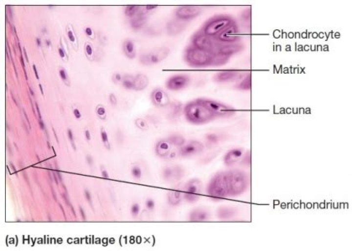
Perichondrium can be found around the perimeter of elastic cartilage and hyaline cartilage. Perichondrium is a type of irregular collagenous ordinary connective tissue, and also functions in the growth and repair of cartilage.
What is the function of the perichondrium quizlet?
It forms the lacunae that surround chondrocytes. It functions in growth and repair of cartilage. It resists outward expansion when cartilage is subjected to pressure. Perichondrium is the type of cartilage that forms the epiglottis.
What is the perichondrium?
Perichondrium is a type of connective tissue, and also functions in the growth and repair of cartilage. Once vascularized, the perichondrium becomes the periosteum. [What type of cartilage has perichondrium?
In elastic cartilage, the chondrocytes are found in a threadlike network of elastic fibres within the matrix. Elastic cartilage provides strength, and elasticity, and maintains the shape of certain structure such as the external ear. It has a perichondrium. This is a diagram of elastic cartilage.
What are Osteoids?
Osteoid is a chemical that the body uses to make bone. It is made by specialized cells called osteoblasts which are found inside bones. When viewed under the microscope, new osteoid looks blue but later turns pink as minerals such as calcium are added to make it stronger.
What is the perichondrium quizlet?
A layer of dense irregular connective tissue. Surrounds the cartilage.
What is perichondrium Class 11?
The perichondrium is a layer of dense irregular connective tissue, which surrounds the cartilage of developing bone. It consists of two separate layers namely, an outer fibrous layer and inner chondrogenic layer. The fibrous layer contains fibroblasts, which produce collagenous fibers.What is Isogenous cell group?
An isogenous group (lat. “equal origin”) is a cluster of up to eight chondrocytes found in hyaline and elastic cartilage.
What is rib perichondrium?Periochondrium means the soft-tissue layer covering the rib cartilage when the rib is exposed. after removal of the surrounding loose soft tissue and the intercostal muscles of the rib. The perichondrium itself is tightly fixed to the underlying rib cartilage. The specific tissue is.
Article first time published onWhat surrounds the epiphysis of the bones to reduce friction?
In this region, the epiphyses are covered with articular cartilage, a thin layer of cartilage that reduces friction and acts as a shock absorber.
What is the hyaline cartilage?
Hyaline cartilage, the most widely distributed form, has a pearl-gray semitranslucent matrix containing randomly oriented collagen fibrils but relatively little elastin. It is normally found on surfaces of joints and in the cartilage making up the fetal skeleton.
What is the function of the deep layer of periosteum?
What is the function of the deep layer of periosteum? Bone remodeling by osteoblasts and osteoclasts.
Is the perichondrium innervated?
Cartilage is an avascular, aneural tissue of the musculoskeletal system. … Owing to the fact that cartilage is an avascular tissue; it obtains its nutrients from adjacent perichondrium that has a rich blood supply, is well innervated and is equipped with a definitive lymphatic system.
What cells are cartilage precursors found in the perichondrium?
- perichondrium – vascularized connective tissue sheath surrounding cartilage (except in case of articular cartilage). …
- chondroblasts – immature cartilage cells.
What is the function of hyaline cartilage?
Articular Cartilage Where bone ends meet to form a joint, they are covered by hyaline cartilage. This cartilage appears bluish white and glistening in a normal healthy joint. Its primary function is to provide some cushioning and minimize friction between the bone ends.
What is it called when blood calcium levels rise above normal bone cells?
The most common cause of high blood calcium is a condition called primary hyperparathyroidism or PHPT. In this condition, one or more of the parathyroid glands produces too much PTH. This, in turn, causes the bones to release too much calcium into the blood.
What is the name of the space that a chondrocyte occupies?
Cartilage. The cartilage cells or chondrocytes are contained in cavities in the matrix, called cartilage lacunae; around these, the matrix is arranged in concentric lines as if it had been formed in successive portions around the cartilage cells. This constitutes the so-called capsule of the space.
Which of the following is not a function of connective tissue?
Absorption of nutrients is not a function of connective tissue.
What are lamellae in bone?
Each osteon consists of concentric layers, or lamellae, of compact bone tissue that surround a central canal, the haversian canal. The haversian canal contains the bone’s blood supplies. … Near the surface of the compact bone, the lamellae are arranged parallel to the surface; these are called circumferential lamellae.
What is the lamellar bone?
Lamellar bone represents the main type of bone in a mature skeleton. It is characterized by an orderly arrangement of collagen bundles and their cells (fig. … The deposited collagen exhibits an orderly lamellar pattern with circular layers of collagen alternating with longitudinal ones.
What is the function of Osteon?
It provides protection and strength to bones. Compact bone tissue consists of units called osteons or Haversian systems. Osteons are cylindrical structures that contain a mineral matrix and living osteocytes connected by canaliculi, which transport blood. They are aligned parallel to the long axis of the bone.
Why Perichondrium is chondrogenic?
The perichondrium consists of an outer fibrous layer that contains fibroblasts and an inner chondrogenic layer that contains chondroblasts. The main functions of the perichondrium are to protect bones from injury and damage, nourish cartilage through blood vessels, and facilitate cartilage growth.
What is the first step in Chondrogenesis?
Each stage was characterized as an established in vitro ESC developmental process of chondrogenesis. As we mentioned previously, the first step in chondrogensis is cell condensation and the subsequent formation of condensed cell aggregates that occurs prior to chondrogenic differentiation [19], [20].
What is Isogenous cell nest?
The two daughter cells produce new matrix and gradually move away from each other. The two daughter cells may divide again before they move away, and we may see two, three or four cells in a single lacuna. Such cell groups are called cell-nests or isogenous groups.
What is the matrix of cartilage called?
Cartilage is composed of specialized cells called chondrocytes that produce a large amount of collagenous extracellular matrix, abundant ground substance that is rich in proteoglycan and elastin fibers. …
What cells are found in the periosteum?
The inner layer of the periosteum contains osteoblasts (bone-producing cells) and is most prominent in fetal life and early childhood, when bone formation is at its peak.
Is calcified cartilage present in human?
In humans, the calcified cartilage is found in the ends of long bones i.e., epiphysis, and in the heads of humerus and femur bone.
Does perichondrium have blood vessels?
Perichondrium. The outer fibrous layer contains blood vessels, lymphatics, and nerves, all of which provide nutrients to, and drain, cartilage.
What type of tissues are the periosteum and perichondrium composed of?
Perichondrium and periosteum are two types of connective tissues present in the body. Perichondrium is a fibrous connective tissue whilst periosteum is a membranous connective tissue. Both connective tissues protect the bones from injury.
How does fibrocartilage get nutrients without perichondrium?
Instead, cartilage relies on two different sources. Synovial Fluid – this is found in joints and supplies nutrients to surrounding chondrocytes (cartilage cells) through diffusion. This is how articular cartilage (the cartilage in bones) receive nutrients, as they don’t have a perichondrium.