What is the inner surface of the eye

Olivia Owen
Published Mar 21, 2026
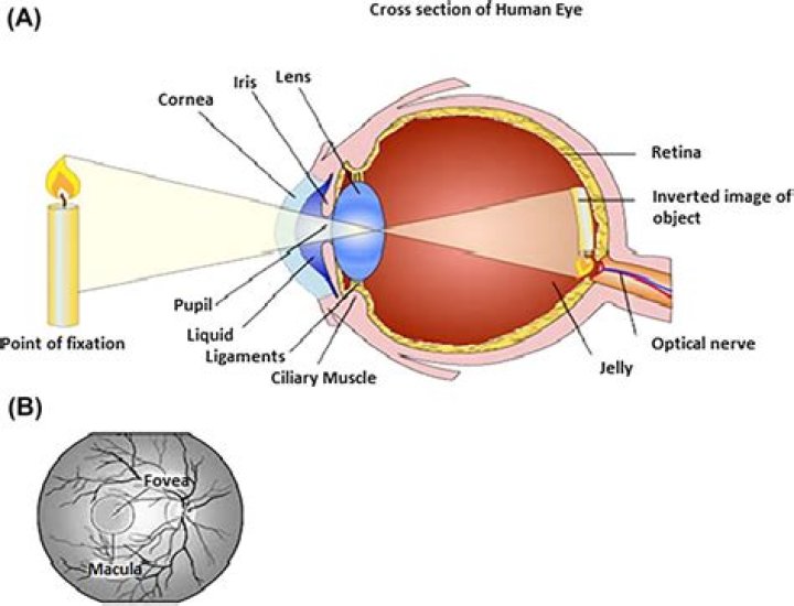
The inner layer is the retina, which lines the back two-thirds of the eyeball.
What is the surface of your eye?
The ocular surface includes the outer layer of the cornea, the tears, the conjunctiva, and the margin of the eye lids. This part of the eye has the most frequent injuries and ocular disease manifestations due to direct exposure to the environment.
Which of the following is the inner layer of the eye which contains rods and cones?
Retina: a light sensitive layer that lines the interior of the eye. It is composed of light sensitive cells known as rods and cones.
What covers the inner surface of the eye and eyelids?
CONJUNCTIVA: The conjunctiva is a transparent mucous membrane that covers the inner surface of the eyelids and the surface of the eye. When it is inflamed or infected it becomes red or pink. This is called conjunctivitis or “pink eye”.Where is the conjunctiva of eye?
It lines the inside of the eyelids and provides a covering to the sclera. It is highly vascularized and home to extensive lymphatic vessels. The conjunctiva can be divided into three regions: the palpebral or tarsal conjunctiva, the bulbar or ocular conjunctiva, and the conjunctival fornices.
Which of the following are mucous membranes that line the inner surfaces of the eyelid?
The conjunctiva is the mucous membrane that lines the eyelid and eye surface. In a healthy eye, the conjunctiva is clear and colourless. The sclera is the tough outer layer of the eyeball (the white of the eye).
What are the layers of the wall of the eye?
The eye is made up of three layers: the outer layer called the fibrous tunic, which consists of the sclera and the cornea; the middle layer responsible for nourishment, called the vascular tunic, which consists of the iris, the choroid, and the ciliary body; and the inner layer of photoreceptors and neurons called the …
What structure contains rods and cones?
Photoreceptor: the special type of cell in your eye that picks up photons and then signals the brain. They are located in the retina (a layer at the back of the eye). There are two types, rods and cones.Where are rods and cones located?
The retina of the eye has two types of light-sensitive cells called rods and cones, both found in layer at the back of your eye which processes images.
Which of these is found in the anterior chamber of the eye?Aqueous humor is the fluid in the anterior chamber of the eye. Vitreous humor is the jellylike substance behind the lens. Remember that aqueous humor is found up front in the anterior cavity of the eye. It provides nourishment to nearby structures and maintains the shape of the anterior eye.
Article first time published onWhich part of the eye is the fluid that the lens floats in?
The vitreous humour (also known simply as the vitreous) is a clear, colourless fluid that fills the space between the lens and the retina of your eye. 99% of it consists of water and the rest is a mixture of collagen, proteins, salts and sugars.
What is conjunctiva and sclera?
The conjunctiva contributes to the tear film and protects the eye from foreign objects and infection. The sclera is the thick white sphere of dense connective tissue that encloses the eye and maintains its shape.
What is the bulbar conjunctiva?
Listen to pronunciation. (BUL-bar kun-JUNK-tih-VY-tis) A condition in which the thin layer of tissue that covers the sclera (the white part of the eye) becomes inflamed. The cause is usually not known, but it may occur with certain inflammatory conditions, such as lupus and rheumatoid arthritis.
What causes pinkeye?
Pink eye is commonly caused by a bacterial or viral infection, an allergic reaction, or — in babies — an incompletely opened tear duct. Though pink eye can be irritating, it rarely affects your vision. Treatments can help ease the discomfort of pink eye.
What is the difference between the rods and cones of the eye?
Rods are responsible for vision at low light levels (scotopic vision). They do not mediate color vision, and have a low spatial acuity. Cones are active at higher light levels (photopic vision), are capable of color vision and are responsible for high spatial acuity.
What is the outer corner of the eye called?
Canthus (pl. canthi, palpebral commissures) is either corner of the eye where the upper and lower eyelids meet. More specifically, the inner and outer canthi are, respectively, the medial and lateral ends/angles of the palpebral fissure.
Which of the following is the inner layer of the eye which contains rods and cones quizlet?
Which of the following is the inner layer of the eye, which contains rods and cones? Iris.
What membrane covers the inside of the eyelid and the anterior white surface of the eye?
The conjunctiva is the clear, thin membrane that covers part of the front surface of the eye and the inner surface of the eyelids. It has two segments: Bulbar conjunctiva. This portion of the conjunctiva covers the anterior part of the sclera (the “white” of the eye).
Which of the following is the beginning of the inner ear?
Vestibular and cochlear systems The footplate of the stapes connects to the oval window, the beginning of the inner ear. When the stapes presses on the oval window, it causes the perilymph, the liquid of the inner ear to move.
What is rod function?
rod, one of two types of photoreceptive cells in the retina of the eye in vertebrate animals. Rod cells function as specialized neurons that convert visual stimuli in the form of photons (particles of light) into chemical and electrical stimuli that can be processed by the central nervous system.
How many rods are in the human eye?
Despite the fact that perception in typical daytime light levels is dominated by cone-mediated vision, the total number of rods in the human retina (91 million) far exceeds the number of cones (roughly 4.5 million). As a result, the density of rods is much greater than cones throughout most of the retina.
What do ganglion cells do?
Retinal ganglion cells process visual information that begins as light entering the eye and transmit it to the brain via their axons, which are long fibers that make up the optic nerve. There are over a million retinal ganglion cells in the human retina, and they allow you to see as they send the image to your brain.
What do cones and rods do in the eye?
Rods and cones are the receptors in the retina responsible for your sense of sight. They are the part of the eye responsible for converting the light that enters your eye into electrical signals that can be decoded by the vision-processing center of the brain.
What is the main function of rods and cones?
Cones and rods are two types of photoreceptors within the retina. This means that they are responsible for receiving signals (or images), processing them, and sending them to the brain.
What is the difference between the anterior and posterior chamber of the eye?
Anterior part of the human eye, with label of posterior chamber at right. … The posterior chamber consists of small space directly posterior to the iris but anterior to the lens. The posterior chamber is part of the anterior segment and should not be confused with the vitreous chamber (in the posterior segment).
What separates the anterior and posterior chambers of the eye?
The iris is the colored portion of your eye. Located behind the cornea, and in front of the crystalline lens. This structure separates the anterior and posterior chambers of the eye. The function of the iris is to help regulate the amount of light that enters your eye.
What is posterior chamber of eye?
Posterior chamber: The posterior chamber is between the iris and lens. The lens is behind the iris and is normally clear. Light passes through the pupil to the lens. The lens is held in place by small tissue strands or fibres (zonules) extending from the inner wall of the eye.
How do floaters form?
Most eye floaters are caused by age-related changes that occur as the jelly-like substance (vitreous) inside your eyes becomes more liquid. Microscopic fibers within the vitreous tend to clump and can cast tiny shadows on your retina. The shadows you see are called floaters.
What is a vitrectomy done for?
A vitrectomy is a type of eye surgery to treat various problems with the retina and vitreous. During the surgery, your surgeon removes the vitreous and replaces it with another solution. The vitreous is a gel-like substance that fills the middle portion of your eye.
Why do I see little balls of light?
Floaters are tiny clumps of cells inside the vitreous (a jelly-like fluid) that fills the inside of the eye. They form as the vitreous gel degenerates, which is part of the normal ageing process. As these cells float in the vitreous gel, they cast shadows on the retina, causing us to see floaters.
What is the difference between the cornea and the conjunctiva?
is that cornea is (anatomy) the transparent layer making up the outermost front part of the eye, covering the iris, pupil, and anterior chamber while conjunctiva is (anatomy) a clear mucous membrane that lines the inner surface of the eyelid and the exposed surface of the eyeball or sclera.