What is the structure of a vesicle

Sarah Rodriguez
Published Feb 23, 2026
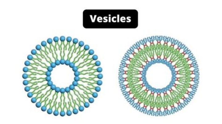
A vesicle is a self-contained structure consisting of fluid or gas surrounded and enclosed by an outer membrane called the lipid bilayer. This is made up of hydrophilic heads and hydrophobic tails that cluster together.
How does the structure of a vesicle affect its function?
The function of vesicles are organelles, and the small enclosed sacs that comprise them can transport and store substances within a cell from one cell to another. They have a lipid bilayer, which separates the contents of the vesicle from the rest of the cell, from the cytoplasm and its contents.
What is the description of vesicle?
Listen to pronunciation. (VEH-sih-kul) A small sac formed by a membrane and filled with liquid. Vesicles inside cells move substances into or out of the cell.
What does a vesicle look like?
Appearance. A typical vesicle looks like a little bubble of fluid under the skin. The larger the vesicle, the more prone to breaking open, which can be quite painful. It can also cause inflammation in the surrounding area.What are the 3 main functions of vesicles?
Vesicles are small cellular containers that perform a variety of functions. They can be used to move molecules, secrete substances, digest materials, or regulate the pressure in the cell.
What is vesicle formation?
In cell biology, a vesicle is a structure within or outside a cell, consisting of liquid or cytoplasm enclosed by a lipid bilayer. Vesicles form naturally during the processes of secretion (exocytosis), uptake (endocytosis) and transport of materials within the plasma membrane.
Which cell structure can form vesicles?
Many vesicles are made in the Golgi apparatus and the endoplasmic reticulum, or are made from parts of the cell membrane by endocytosis. Vesicles can also fuse with the cell membrane and release their contents to the outside.
Where are vesicles located in a neuron?
Explanation: Synaptic vesicles are located in the axon terminals (in the synaptic bulbs), close to the presynaptic membrane ready to deliver the neurotransmitters by exocytosis.Do vesicles have membranes?
A vesicle is a self-contained structure consisting of fluid or gas surrounded and enclosed by an outer membrane called the lipid bilayer.
What is vesicle fluid?Vesicles are small fluid-filled sacs or blisters that can appear on your skin. The fluid inside these sacs may be clear, white, yellow, or mixed with blood. Vesicles are fluid-filled lesions less than 5 mm (1/2 cm). If the fluid-filled lesion is greater than 0.5 mm, it’s called a bulla.
Article first time published onHow do vesicles move in a cell?
In general, vesicles move from the ER to the cis Golgi, from the cis to the medial Golgi, from the medial to the trans Golgi, and from the trans Golgi to the plasma membrane or other compartments. … When associated with transmembrane proteins, they can pull the attached membrane along into a spherical shape also.
What structures are present in an animal cell but not in a plant cell?
Animal Cells versus Plant Cells Animal cells each have a centrosome and lysosomes, whereas plant cells do not. Plant cells have a cell wall, chloroplasts and other specialized plastids, and a large central vacuole, whereas animal cells do not.
What is a vesicle in pathology?
Vesicles are small, circumscribed, subcorneal (intraepidermal) or subepidermal cystlike spaces that contain serous fluid ( Figure 1. ). They represent detachment of damaged epithelium with the resulting space being filled by fluid.
What is the function of vesicles in the synthesis of proteins?
Vesicles transport the proteins from the ribosomes to the Golgi apparatus, a.k.a Golgi complex, where they are packaged into new vesicles. The vesicles migrate to the membrane and release their protein to the outside of the cell. Lysosomes digest and recycle the waste materials for reuse by the cell.
Are vesicles prokaryotic or eukaryotic?
Eukaryotic CellProkaryotic CellVesiclesPresentPresentGolgi ApparatusPresentAbsentMitosisYesNo; binary fissionChloroplastsPresent in plantsAbsent; chlorophyll is scattered in the cytoplasm
Which structure controls the processes of the cell?
The nucleus, formed by a nuclear membrane around a fluid nucleoplasm, is the control center of the cell.
In which of the following does the membrane of a vesicle become part of the cell membrane?
When a secretory vesicle fuses with the plasma membrane, its contents are discharged from the cell by exocytosis, and its membrane becomes part of the plasma membrane.
Can cell surface membrane form vesicles?
At present, evidence of two major pathways is found. One is that the vesicle is formed on the membrane surface, either at the plasma membrane at the cell surface or at internal cell organelles like the TGN. Another possibility is that vesicles formed elsewhere dock at, fuse with, and then break off from membranes.
What is structure of cell wall?
The main functions of the cell wall are to provide structure, support, and protection for the cell. The cell wall in plants is composed mainly of cellulose and contains three layers in many plants. The three layers are the middle lamella, primary cell wall, and secondary cell wall.
How do you make vesicles?
- Dissolve the lipids in chloroform.
- Combine the lipids in the appropriate ratio.
- Carefully evaporate the organic solvent using a dry nitrogen stream.
- Resuspend the lipid mixture in cyclohexane. …
- Freeze the cyclohexane solution using dry ice.
How do vesicles form in igneous rocks?
As magma rises to the surface the pressure on it decreases. … When the magma finally reaches the surface as lava and cools, the rock solidifies around the gas bubbles and traps them inside, preserving them as holes filled with gas called vesicles.
How does the structure of the membrane allow the formation of vesicles?
The phospholipids in the cell membrane are not solid but are in a fluid state allowing the membrane to change its shape and also vesicles to fuse with it. This means substances can enter the cell via endocytosis and exit the cell via exocytosis. The membrane then returns to its original state.
How do microtubules form?
Microtubules are polymers of tubulin that form part of the cytoskeleton and provide structure and shape to eukaryotic cells. … They are formed by the polymerization of a dimer of two globular proteins, alpha and beta tubulin into protofilaments that can then associate laterally to form a hollow tube, the microtubule.
Where is the secretory vesicle located?
a. They are located at presynaptic terminals in neurons. When a signal reaches the end of an axon, the synaptic vesicles fuse with the cell membrane and release the neurotransmitter.
What part of the neuron releases neurotransmitters from vesicles?
The axonal terminals are specialized to release the neurotransmitters of the presynaptic cell. * Neurotransmitter molecule-stored in small “packages” called vesicles, released from the axon terminal when their vesicles “fuse” with the membrane of the axon terminal, spilling the neurotransmitter into the synaptic cleft.
How do synaptic vesicles move?
To move synaptic vesicles within the nerve terminal environment, tethering proteins interact to chaperone (aid in the movement of) vesicles between storage pools, recycling pools, and the docking sites for transmitter release (see Chapter 9, for more information on these synaptic vesicle pools).
What is the basic structural and functional unit of the nervous system?
Neurons are the basic functional units of the nervous system, and they generate electrical signals called action potentials, which allow them to quickly transmit information over long distances. Glia are also essential to nervous system function, but they work mostly by supporting the neurons.
What are vesicles and bullae?
Vesicles and. Bullae: (Blisters) Vesicles are circumscribed epidermal elevations in the skin containing clear fluid and less than ½ cm. in diameter. If the lesion has a diameter of greater than ½ cm, it is called a bulla. Vesicles and bullae arise from a cleavage at various levels of the skin.
How do vesicles fuse with the membrane?
These two proteins may allow the vesicle and presynaptic membrane to recognize each other. Following docking, there is a second influx of calcium at the active zone, which causes the vesicle membrane to fuse to the presynaptic membrane, forming a temporary ion channel.
What structures are found in all cells?
All cells share four common components: 1) a plasma membrane, an outer covering that separates the cell’s interior from its surrounding environment; 2) cytoplasm, consisting of a jelly-like region within the cell in which other cellular components are found; 3) DNA, the genetic material of the cell; and 4) ribosomes, …
What is structures that converts nutrients to energy?
Mitochondria – organelles that, using oxygen, convert nutrients into energy that can be used by the cell.