What is the zygomatic buttress

Andrew Campbell
Published May 14, 2026
the structural pillar of the mid-face that extends superiorly from the maxillary ridge through the zygomatic bone to the frontal and temporal bones.
What does the zygomatic do?
The zygomatic bone functions as a structure which joins the bones of the face while protecting the arteries, nerves, veins, and organs which lie below the surface. The arches of the zygomatic bone provide a person’s cheeks with the structure to fill out the face.
How many mandibular buttresses are there?
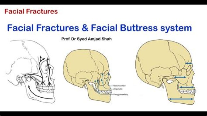
The four horizontal buttresses are the upper transverse maxillary buttress, lower transverse maxillary buttress, upper transverse mandibular buttress, and lower transverse mandibular buttress [30]; the frontal bar could be included as a fifth buttress [31].
What does zygomatic mean?
Definition of zygomatic : of, relating to, constituting, or situated in the region of the zygomatic bone or zygomatic arch.What is the zygomatic arch in dentistry?
With the use of a zygomatic dental implant, it is placed directly to the upper jawbone (zygomatic arch) which is a dense facial bone that can provide stable support to implants.
What bones make lower jaw?
The mandible is the largest bone in the human skull. It holds the lower teeth in place, it assists in mastication and forms the lower jawline. The mandible is composed of the body and the ramus and is located inferior to the maxilla. The body is a horizontally curved portion that creates the lower jawline.
What is Zygomaticus major?
Of all the muscles in the face, the zygomaticus major is perhaps the most noticeable. Sitting between the corners of our lips and the upper part of our cheeks, it controls the way in which we smile. The muscle sits atop the zygomatic bone, otherwise known as the cheekbone.
What is another name for zygoma?
zygomatic bone, also called cheekbone, or malar bone, diamond-shaped bone below and lateral to the orbit, or eye socket, at the widest part of the cheek.What two bones make up the zygomatic arch?
The cranial portion of the zygomatic arch is formed by the zygomatic bone, and the caudal portion is formed by the zygomatic process of the temporal bone. The zygomatic arch forms the ventral and lateral rim of the orbit.
Why is it called the zygoma?Zygomatic bone: The part of the temporal bone of the skull that forms the prominence of the cheek. … The word “zygomatic” comes from the Greek “zygon” meaning a yoke or crossbar by which two draft animals such as oxen could be hitched to a plow or wagon.
Article first time published onWhat is buttress anatomy?
buttress. (lip’ō-oks’ĭ-jen-ās), A structure placed against the base of another to support or stabilize it.
Where is the zygomatic buttress?
the structural pillar of the mid-face that extends superiorly from the maxillary ridge through the zygomatic bone to the frontal and temporal bones.
What is maxillofacial trauma?
Maxillofacial trauma is any injury to the face or jaws. Facial trauma may present with skin lacerations, burns, obstruction to the nasal cavity or sinuses, damage to the orbital (eye) sockets, fracture to the jawbone, and missing or broken teeth.
How successful are zygomatic implants?
Like traditional dental implants, zygomatic dental implants also have a high success rate. It is estimated that around the success rate of zygomatic implants ranges from 97-98%. In the case of complications, the most common complications of zygomatic implants are sinusitis, paresthesia, and oro-antral fistula.
What can I eat after zygomatic implants?
You can eat anything that you can cut with a fork such as meatloaf, shredded chicken, scrambled eggs, cooked vegetables, mashed potatoes, etc. If you can hear yourself chewing, it’s too hard. Remember, although your gum tissue will appear healed in about 10-14 days, the implants will NOT be strong for 5 months.
Do zygomatic implants fail?
A total of 56 zygomatic implants were reported as failures and the cumulative success rate (CSR) over a 12-year period was 96.7% (6). The preliminary data show that the zygomatic implant technique is predictable with satisfactory clinical outcomes.
What facial muscle is used in smiling?
A, the orbicularis oculi and zygomaticus major muscles during smiling.
How many muscles used smiling?
About 43 muscles in a face are working to create a smile at any given moment. It is proven by Dr. Ekman’s research tool called FACS or Facial Action Coding System.
Does smiling strengthen cheek muscles?
Smile as widely as you can and press your fingertips into the folds between your nose and lips. Lift up the muscles whilst pressing down your fingertips into the muscles for resistance. This will strengthen your cheek muscles to give you plump, round cheeks – a classic sign of youth.
Do humans have two jaws?
In the human fetus and infant both the upper and lower jaws have two halves; these fuse at the midline a few months after birth.
Are teeth bones?
Even though teeth and bones seem very similar, they are actually different. Teeth are not bones. Yes, both are white in color and they do indeed store calcium, but that’s where their similarities end.
What is the name of the bone that houses the brain?
The cranium (skull) is the skeletal structure of the head that supports the face and protects the brain. It is subdivided into the facial bones and the brain case, or cranial vault (Figure 6.16).
What muscles attach to the Zygoma?
- Zygomaticus major.
- Zygomaticus minor.
- Masseter.
- Lateral palpebrae ligament (part of the levator palpebrae superioris)
- Levator labii superioris (the origin is the maxillary border of the zygomatic bone)
Does a crocodile have a zygomatic arch?
The crocodile, for instance, has no zygomatic arch and has one of the most powerful bites in the animal kingdom. Using the zygomatic arch for bite comparisons is really only useful between animals that evolved with a zygomatic arch, as other animals use different bones for similar purposes.
What is the weakest part of the skull?
Clinical significance The pterion is known as the weakest part of the skull. The anterior division of the middle meningeal artery runs underneath the pterion. Consequently, a traumatic blow to the pterion may rupture the middle meningeal artery causing an epidural haematoma.
What does zygomatic bone articulate with?
The zygomatic bone articulates with the sphenoid bone, maxilla, frontal bone, and temporal bone to form the lateral wall of the floor of the orbit, part of the temporal and infratemporal fossa, and the prominence of the cheek.
How do you pronounce zygoma?
noun, plural zy·go·ma·ta [zahy-goh-muh-tuh, zi-]. Anatomy.
What is another name for collar bone?
clavicle, also called collarbone, curved anterior bone of the shoulder (pectoral) girdle in vertebrates; it functions as a strut to support the shoulder.
What chewing muscle attaches on the zygomatic arch?
The masseter muscle, important in chewing, arises from the lower edge of the arch; another major chewing muscle, the temporalis, passes through the arch. The zygomatic arch is particularly large and robust in herbivorous animals, including baboons and apes.
What are the cheekbones?
Your cheekbones are the structure of your face underneath your skin, particularly the malar bones. If your malar bones are located in close proximity to your eyes, you’re considered to have high cheekbones. Lower cheekbones refer to malar bones that rest closer to the bottom of your nose.
What is the anatomical term for cheek?
CheekFMA46476Anatomical terminology