Where does CN III exit the brainstem

Sarah Rodriguez
Published Apr 29, 2026
The oculomotor nerve
Where does each cranial nerve exit the brain?
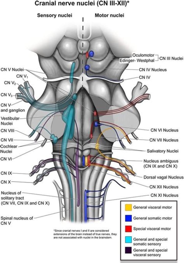
The cranial nerves are nerves that arise from the brain and exit the skull through holes (cranial foramina) at its base rather than through the spinal cord.
Where does the Trochlear nerve terminate?
The trochlear nerve terminates within the posterior half of the superior oblique muscle (Figure 13); in 76% of the orbits it ends in the medial aspect of the muscle and in the remaining 24% it ends in the superior aspect of the muscle.
What foramen does the oculomotor nerve pass through?
Cranial NerveForamenComponentsII-Opticoptic canal of sphenoidspecial sensoryIII-Oculomotorsuperior orbital fissuresomatomotorvisceromotorIV-Trochlearsuperior orbital fissuresomatomotorWhat is the main function of CN I?
CNs have sensory (afferent) and motor (efferent) functions. CN I and CN II convey sensory information. … All the other CNs originate from the brainstem nuclei (the hypoglossal nerve is located at the border of the spinal cord) and include sensory axons as well as motor axons.
What is the function of CN III V VII to eyelids?
Motor. Eyelid muscle innervation is achieved by cranial nerve VII (the facial nerve), cranial nerve III (the oculomotor nerve), and sympathetic nerve fibers. The facial nerve (CNVII) innervates the orbicularis oculi, frontalis, procerus, and corrugator supercilii muscles, and supports eyelid protraction.
Which cranial nerve exits the dorsal aspect of the brainstem?
The Trochlear Nerve (IV) It decussates at the anterior medullary velum in the roof of the aqueduct before exiting from dorsal midbrain below the inferior colliculus (Fig. 4.7). The nerve is the only motor cranial nerve that leaves from the dorsal side of the brainstem.
How do you test for CN 3?
Inability to follow and object in direction of CN III (the quickest test is to observe upward gaze which is all CN III; the eye on the affected side does not look upward) Inability to open the eyelid. CN III dysfunction causes the eyelid on the affected side to become “droopy”. This is called ptsosis.Which opening in the occipital bone allows the hypoglossal nerve CN XII to exit the skull?
The hypoglossal nerve leaves the skull through the hypoglossal canal, which is situated near the large opening for the spinal cord, the foramen magnum. After leaving the skull, the hypoglossal nerve spirals around the vagus nerve and then passes behind the deep belly of the digastric muscle.
What is CN IV?This nerve is the fourth set of cranial nerves (CN IV or cranial nerve 4). It is a motor nerve that sends signals from the brain to the muscles. CN IV works with the oculomotor nerve and other eye muscles to control eye movement.
Article first time published onWhat is CN IV palsy?
Fourth nerve palsy means that a certain muscle in your eye is paralyzed. It is caused by disease or injury to the fourth cranial nerve. In children, it is most often present at birth (congenital). In adults, it is most often caused by injury. Many cases of fourth nerve palsy are idiopathic.
What is special about CN IV?
CNIV is unique in that it has a long path to its origin and is the only cranial nerve that exits the brainstem dorsally (towards the back). … The long pathway, as well as the dorsal exit of the nerve, makes this nerve one of the most susceptible to damage from a head injury.
What is the main function of CN III?
The oculomotor nerve is the third cranial nerve (CN III). It allows movement of the eye muscles, constriction of the pupil, focusing the eyes and the position of the upper eyelid. Cranial nerve III works with other cranial nerves to control eye movements and support sensory functioning.
What is the main function of CN XII quizlet?
CN XII is responsible for tongue movement. CN XI is responsible for neck and shoulder movement. CN IX is responsible for taste in the posterior two thirds of the tongue, pharyngeal sensation, and swallowing.
What is the main function of CN VIII quizlet?
What is the function of cranial nerve VIII? The vestibulocochlear nerve is responsible for hearing and equilibrium.
Which cranial nerve innervates the posterior 1/3 of the tongue?
On the other hand, taste to the posterior one-third of the tongue is accomplished through innervation from the glossopharyngeal nerve (CN IX), which also provides general sensation to the posterior one-third of the tongue.
How do you test for CN IV?
Trochlear nerve (CN IV) Cranial nerve IV acts as a pulley to move the eyes down—toward the tip of the nose. To assess the trochlear nerve, instruct the patient to follow your finger while you move it down toward his nose.
Where does the optic nerve exit from the retina?
At the optic disc, the optic nerve fibers exit the eye through fenestrations within the sclera, termed the lamina cribrosa. The retinal ganglion cell axons remain unmyelinated until traversing the lamina cribrosa.
What nerve closes the eye?
The facial nerve is responsible for closing the eyes by contracting the orbicularis oculi muscle.
What nerve controls left eyelid?
The oculomotor nerve is the third of 12 pairs of cranial nerves in the brain. This nerve is responsible for eyeball and eyelid movement. It follows the olfactory and optic nerves in terms of order. The oculomotor nerve involves two separate components, each of which has a distinct function.
Which cranial nerve is CN VII?
The facial nerve is the seventh cranial nerve (CN VII). It arises from the brain stem and extends posteriorly to the abducens nerve and anteriorly to the vestibulocochlear nerve.
Where does the hypoglossal nerve terminate?
It’s also known as the 12th cranial nerve, cranial nerve 12 or CNXII. This nerve starts at the base of your brain. It travels down your neck and branches out, ending at the base and underside of your tongue.
Where does the accessory nerve exit the skull?
The nerve travels along the inner wall of the skull towards the jugular foramen. Leaving the skull, the nerve travels through the jugular foramen with the glossopharyngeal and vagus nerves. The spinal accessory nerve is notable for being the only cranial nerve to both enter and exit the skull.
What happens if cranial nerve 3 is damaged?
Third cranial nerve disorders can impair ocular motility, pupillary function, or both. Symptoms and signs include diplopia, ptosis, and paresis of eye adduction and of upward and downward gaze. If the pupil is affected, it is dilated, and light reflexes are impaired.
Which muscle is innervated by the Trochlear N CN IV )?
The only muscle the trochlear nerve innervates, the superior oblique muscle, is the longest and thinnest muscle among the extraocular muscles.
What is the location of the trigeminal nerve?
Each trigeminal ganglion is located near your temple at the side of your head, in front of your ear. The trigeminal ganglion splits into three trigeminal nerve branches. These branches travel along each side of your head to different parts of your face.
What is Hypertropia of the eye?
A hypertropia is a form of vertical strabismus where one eye is deviated upwards in comparison to the fellow eye. The term of hypertropia is relative to the fellow eye which, by analogy is the hypotrpoic eye- meaning that is deviated downwards.
How common is 4th cranial nerve palsy?
According to a study conducted by the American Academy of Ophthalmology (AAO), the most common type of fourth nerve palsy is congenital (49%), followed by hypertension (18%) and trauma (18%). Concussions and whiplash are the most common causes of injury to the fourth cranial nerve.
What causes Trochlear nerve palsy?
The most common cause of congenital trochlear nerve palsies is congenital cranial dysinnervation syndrome, followed by an abnormal superior oblique tendon. The most common cause of acquired isolated fourth nerve palsy, after idiopathic, is head trauma.
How does the abducens nerve move the eye?
The abducens nerve leaves the brainstem at the junction of the pons and the medulla, medial to the facial nerve. It runs upwards and forwards from this position to reach the eye. … It then enters the orbit through the superior orbital fissure and innervates the lateral rectus muscle of the eye.
Where is the Interpeduncular fossa?
The interpeduncular fossa is a somewhat rhomboid-shaped area of the base of the brain, limited in front by the optic chiasma, behind by the antero-superior surface of the pons, antero-laterally by the converging optic tracts, and postero-laterally by the diverging cerebral peduncles.