Where is lumbosacral plexus

Olivia Owen
Published May 05, 2026
The lumbosacral plexus is a network of nerves derived from lumbar and sacral roots with each one of them dividing into anterior and posterior branches. Their communications are called lumbar plexus
Where is the lumbosacral plexus located?
The lumbosacral plexus is formed by the anterior rami of the nerves (spinal segments T12–S4) to supply the lower limbs. The lumbosacral plexus can be divided into the lumbar plexus, which innervates the ventral upper half, and the sacral plexus, which mainly innervates the dorsal side.
Where are the plexuses located?
Nerve Junction Boxes: The Plexuses Four nerve plexuses are located in the trunk of the body: The cervical plexus provides nerve connections to the head, neck, and shoulder. The brachial plexus provides connections to the chest, shoulders, upper arms, forearms, and hands.
Where does the lumbosacral plexus originate?
Origin and location The lumbar plexus originates from the anterior rami of spinal nerves L1-L4 and is formed largely within the posterior aspect of the psoas major muscle.What is a lumbosacral?
Of or relating to or near the small of the back and the back part of the pelvis between the hips. The lumbosacral junction consist of the L5 vertebral body articulating with the first sacral vertebral body.
What nerve is associated with the lumbosacral plexus?
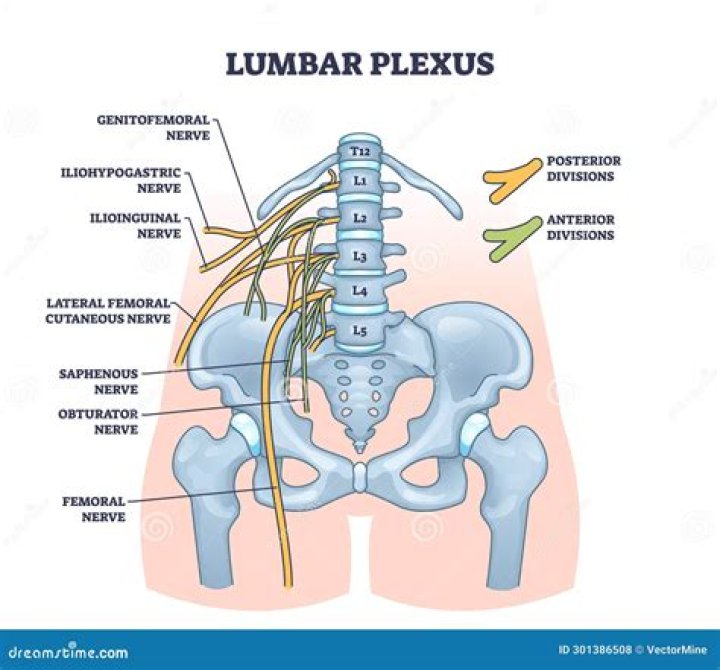
The nerves arising from the lumbar plexus from superior to inferior are iliohypogastric, ilioinguinal, genitofemoral, lateral femoral cutaneous, femoral nerve, obturator, and nerve to the lumbosacral trunk.
What is a lumbosacral plexus injury?
The lumbosacral plexus is a network of nerve fibres supplying the muscles of your lower limbs. It’s located in your lower back, and forks into branches in front of the hip joint and down your legs. Damage, at birth or due to a trauma, to the lumbosacral plexus causes pain and weakness in the lower back.
What is the meaning of plexuses?
Definition of plexus 1 : a network of anastomosing or interlacing blood vessels or nerves. 2 : an interwoven combination of parts or elements in a structure or system.What is the cauda?
Cauda is Latin for tail, and equina is Latin for horse (ie, the “horse’s tail”). The CE provides sensory innervation to the saddle area, motor innervation to the sphincters, and parasympathetic innervation to the bladder and lower bowel (ie, from the left splenic flexure to the rectum).
How are plexuses formed?A nerve plexus is a network of intersecting nerves; multiple nerve plexuses exist in the body. Nerve plexuses are composed of afferent and efferent fibers that arise from the merging of the anterior rami of spinal nerves and blood vessels.
Article first time published onWhat are the 4 nerve plexuses?
Of the four major nerve plexuses (cervical, brachial, lumbar, and sacral), only the brachial plexus and sacral plexus can be assessed satisfactorily in the EDX laboratory.
What vertebrae are lumbosacral?
The spine is composed of 33 interlocking bones called vertebrae. The lumbosacral region of the spine consists of 5 lumbar vertebrae and the sacrum (5 bones joined together).
What is considered the lumbosacral region?
The sacral region (sacrum) is at the bottom of the spine and lies between the fifth segment of the lumbar spine (L5) and the coccyx (tailbone).
What is the difference between lumbar region and lumbosacral region?
As adjectives the difference between lumbar and lumbosacral is that lumbar is related to the lower back or loin while lumbosacral is of or pertaining to the lumbar and sacral regions of the back; the small of the back and the back portion of the pelvis.
What happens if the cervical plexus is damaged?
Damage to the cervical plexus can cause sensory disturbances to the posterior head, neck, submandibular region, and the superior back, in a cape-like distribution.
What does the sacral plexus do?
The sacral plexus (plexus sacralis) is a nerve plexus that provides motor and sensory nerves for the posterior thigh, most of the lower leg, the entire foot, and part of the pelvis (see the following image).
Is lumbar spine the same as lumbosacral spine?
A lumbosacral spine x-ray is a picture of the small bones (vertebrae) in the lower part of the spine. This area includes the lumbar region and the sacrum, the area that connects the spine to the pelvis. This is the spine and the sacrum with the cervical (neck), thoracic (mid-back), and lumbar (lower back) vertebra.
What are the first signs of cauda equina?
- Lower limb weakness and intermittent changes in sensation, such as numbness.
- “Saddle anesthesia” – loss or diminished sensation in areas where a person would sit on a saddle.
- Urinary and/or bowel problems, such as retention or incontinence.
What is phylum terminal?
The filum terminale (FT) is a fibrous band that extends from the conus medullaris to the periosteum of the coccyx, and its functions are to fixate, stabilize, and buffer the distal spinal cord from normal and abnormal cephalic and caudal traction.
What is saddle anesthesia?
Saddle anaesthesia refers to reduced sensation in the area that would be in contact with a saddle if sitting on one. This includes the perineum, buttocks, anus, groin and upper thighs. Saddle anaesthesia will make these areas feel numb and abnormal.
How many plexuses are in the body?
Plexuses. The four primary nerve plexuses are the cervical plexus, brachial plexus, lumbar plexus, and the sacral plexus.
What is cervical plexus?
The cervical plexus is a plexus of the anterior rami of the first four cervical spinal nerves which arise from C1 to C4 cervical segment in the neck. They are located laterally to the transverse processes between prevertebral muscles from the medial side and vertebral (m. scalenus, m.
What makes up the pulmonary plexus?
The pulmonary plexus is an autonomic plexus formed from pulmonary branches of vagus nerve and the sympathetic trunk. The plexus is in continuity with the deep cardiac plexus.
What are autonomic plexuses?
autonomic plexus: Any of the extensive networks of nerve fibers and cell bodies associated with the autonomic nervous system that are found in the thorax, abdomen, and pelvis, and that contain sympathetic, parasympathetic, and visceral afferent fibers.
Which Rami form the plexuses?
Ventral ramusTA26147FMA5982Anatomical terms of neuroanatomy
What is lumbosacral or cervical strain?
Lumbosacral or cervical strain is an injury of the ligaments, tendons and/or muscles of the low back or neck, respectively. The injury usually results from stretching that causes a small tear in these tissues. Lumbosacral and cervical strain are typically caused by overuse and trauma.
Where is lumbar 3 and 4?
The L3-L4 spinal motion segment, positioned in the middle of the lumbar spine, plays an important role in supporting the weight of the torso and protecting the cauda equina (nerves that descend from the spinal cord).
Where is vertebrae 4 and 5?
The L4 and L5 are the two lowest vertebrae of the lumbar spine. Together with the intervertebral disc, joints, nerves, and soft tissues, the L4-L5 spinal motion segment provides a variety of functions, including supporting the upper body and allowing trunk motion in multiple directions.
What does sacroiliac joint pain feel like?
You may experience sacroiliac (SI) joint pain as a sharp, stabbing pain that radiates from your hips and pelvis, up to the lower back, and down to the thighs. Sometimes it may feel numb or tingly, or as if your legs are about to buckle.