Why are the bones of a fetal skull not ossified

Olivia Owen
Published Mar 23, 2026
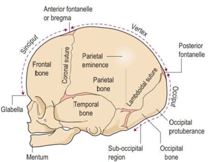
The bones of the newborn skull are not fully ossified and are separated by large areas called fontanelles, which are filled with fibrous connective tissue. … At the time of birth, the facial bones are small and underdeveloped, and the mastoid process has not yet formed.
Why is the skull not fully ossified at birth?
The bones of the newborn skull are not fully ossified and are separated by large areas called fontanelles, which are filled with fibrous connective tissue. … At the time of birth, the facial bones are small and underdeveloped, and the mastoid process has not yet formed.
When do fetal bones ossify?
Bone ossification, or osteogenesis, is the process of bone formation. This process begins between the sixth and seventh weeks of embryonic development and continues until about age twenty-five; although this varies slightly based on the individual.
Why are the bones of the fetal skull not fused?
When a baby has craniosynostosis, one or more of the sutures closes too early, and the baby’s skull continues growing in an abnormal fashion. Sometimes the brain gets squeezed by the skull bones that fused too soon, which can cause pressure to build up inside of the skull.When does the skull become completely ossified?
The fully formed adult human skull is formed from fused skull bones, with all remaining soft spots covered with expanding cranial bone. Although at this stage, it is considered a “full grown” skull, the seams between the bones of the skull do not completely fuse together until about age 20.
What does not articulate with any other bone?
Famously, the hyoid bone is the only bone in humans that does not articulate with any other bone, but only has muscular, ligamentous, and cartilaginous attachments.
Which of the following bones is not a part of the appendicular skeleton?
The hyoid is a bone located in the throat, and it is the only bone in our body that is not connected to another bone. The main function of the human skeleton is to protect our internal organs and provide support and attachment surfaces for our muscles to make the body mobile.
Why is it important for the skull to have holes through the bones?
A foramen allows a structure to pass through, so things like blood vessels and nerves. In the skull, they’re really important to know because many important blood vessels pass through these foramina to supply the brain and several important nerves pass through them to carry information to and from the brain.Why do you think the curved bones of the skull are fused together?
This means that the skull can flex and deform during birth, making it easier to deliver a baby through the narrow birth canal. These individual plates of bone fuse together after about 24 months to form the adult skull.
When does the skull fuse on a baby?The sutures of the skull fuse around the brain at around age 2 years. When a baby has craniosynostosis, one or more of these sutures hardens too early and closes before the baby reaches age 2.
Article first time published onHow does bone ossify?
Time periodBones affectedThird month of fetal developmentOssification in long bones beginning
What happens as bones ossify quizlet?
What happens in Intramembranous Ossification? Osteoblasts become trapped / convert to osteocytes. Bone growth continues / forms ossification centers (spicules). Blood vessels branch into region between spicules.
How does ossification occur in the embryo?
endochondral ossification: A process that occurs during fetal development by which bone tissue is created using a cartilage template. … The membrane that occupies the place of the future bone resembles connective tissue and ultimately forms the periosteum, or outer bone layer.
What do you call the areas of membrane that have not yet ossified in the fetus or newborn?
The spaces between the bones that remain open in babies and young children are called fontanelles. Sometimes, they are called soft spots. These spaces are a part of normal development.
What is the difference between neurocranium and viscerocranium?
The adult human skull consists of two regions of different embryological origins: the neurocranium and the viscerocranium. The neurocranium is a protective shell surrounding the brain and brain stem. The viscerocranium (or facial skeleton) is formed by the bones supporting the face.
What are the causes of abnormalities in skull size?
Most skull deformities result from abnormal development of the brain or from premature closure of some sutures. Babies born with acrania (absence of calvaria) fail to survive because most of the brain is absent. In microcephaly, the size of the brain is very small, and consequently the skull fails to grow.
What is appendicular bone?
The appendicular skeleton is one of two major bone groups in the body, the other being the axial skeleton. The appendicular skeleton is comprised of the upper and lower extremities, which include the shoulder girdle and pelvis.
Is skull axial or appendicular?
The axial skeleton includes the bones that form the skull, laryngeal skeleton, vertebral column, and thoracic cage. The bones of the appendicular skeleton (the limbs and girdles) “append” to the axial skeleton.
What makes up the appendicular skeleton?
The appendicular skeleton includes the bones of the shoulder girdle, the upper limbs, the pelvic girdle, and the lower limbs.
Which one of the following is au shaped bone and which does not articulate with other bones?
Information. The hyoid bone in the neck is the only bone in the body that does not articulate directly with at least one other bone. It is U-shaped and is held in place by, and helps anchor, muscles that connect to the floor of the mouth and the tongue.
What bone does not articulate with any other bones quizlet?
The hyoid bone is the only bone of the body that does not articulate with any other bone.
Which of the following bones is not part of the skull?
The maxilla is the only listed bone that is not part of the cranium. Instead, it is a facial bone.
Which of the following bones is not part of the orbit of the skull ethmoid sphenoid Vomer frontal?
Which of the following bones does NOT form a part of the orbits of the eyes? Seven bones contribute to the orbits. They are the frontal, sphenoid, zygomatic, maxilla, palatine, lacrimal, and ethmoid bones. The vomer is not a contributor to the orbits.
Which of the following is not a function of the facial bones of the skull?
Which of the following is NOT a function of the facial bones of the skull? The cranial cavity, which does protect the brain, is not made up of facial bones.
Which of the following bones is not a facial bone quizlet?
True. ETHMOID BONE is one of eight CRANIAL BONES, not the facial bone!
What goes through the holes in the skull?
The human skull has numerous foramina through which cranial nerves, arteries, veins, and other structures pass. The skull bones that contain foramina include the frontal, ethmoid, sphenoid, maxilla, palatine, temporal, and occipital lobes.
In which type of fracture does the bone completely break but does not pierce through the skin?
A closed fracture is when the bone breaks but there is no puncture or open wound in the skin. An open fracture is one in which the bone breaks through the skin; it may then recede back into the wound and not be visible through the skin.
What are the main functions of the skull?
The main function of the bones of the skull along with the surrounded meninges, is to provide protection and structure. Protection to the brain (cerebellum, cerebrum, brainstem) and orbits of the eyes.
Why is there a dip in my baby's head?
It is normal for a fontanel to form an inward curve in infants while their skull is still hardening. But in some cases, it may become sunken, and the cause may need medical treatment. A sunken fontanel, when accompanied by other symptoms, can be a sign of dehydration or malnutrition.
What is Pfeiffer syndrome?
Pfeiffer syndrome, also known as acrocephalosyndactyly Type V, is a genetic disorder characterized by the anomalies of the skull, face and limbs. Gene mutations are responsible for causing the early fusion of the skull, hand and feet bones. Craniofacial differences are similar to those seen in Apert syndrome.
What does a sunken fontanel look like?
The one on the top of the head remains present until your baby is between 7 and 19 months old. A baby’s soft spots should be relatively firm and curve ever so slightly inward. A soft spot with a noticeable inward curve is known as a sunken fontanel.