What does the T wave represent on an ECG

Olivia Owen
Published Mar 11, 2026
Introduction. The T wave on the ECG (T-ECG) represents repolarization of the ventricular myocardium. Its morphology and duration are commonly used to diagnose pathology and assess risk of life-threatening ventricular arrhythmias.
What does end of T wave represent in ECG?
In electrocardiography, the T wave represents the repolarization of the ventricles. The interval from the beginning of the QRS complex to the apex of the T wave is referred to as the absolute refractory period. The last half of the T wave is referred to as the relative refractory period or vulnerable period.
Where is the T wave on an ECG?
The T wave occurs after the QRS complex and is a result of ventricular repolarization. T waves should be upright in most leads; the exceptions are aVR and V1. Further, T waves should be asymmetric in nature.
What happens in the T wave?
The T wave represents ventricular repolarization. Generally, the T wave exhibits a positive deflection. The reason for this is that the last cells to depolarize in the ventricles are the first to repolarize.What each ECG wave represents?
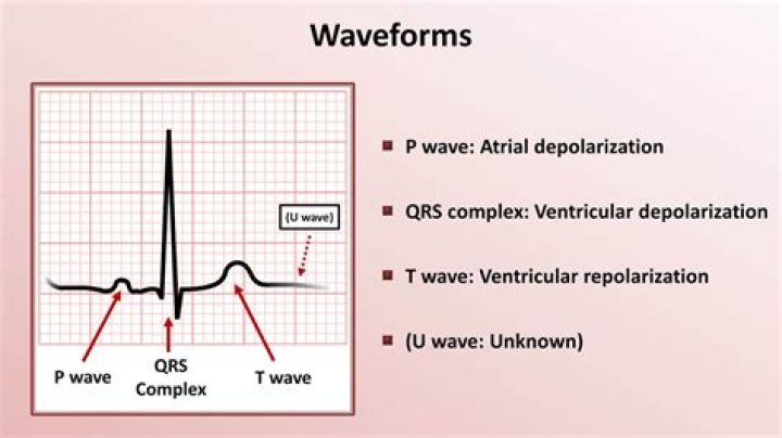
There are three main components to an ECG: the P wave, which represents the depolarization of the atria; the QRS complex, which represents the depolarization of the ventricles; and the T wave, which represents the repolarization of the ventricles.
What does a notched T wave mean?
A notched T-wave was defined as the presence of a bifid T-wave with a notch duration between the 2 peaks ≥0.04 sec and an amplitude ≥0.05 mV. We excluded misinterpretations with prominent U-waves fused to the end of the T-wave or with hidden P-waves embedded in the T-wave.
What do tall T waves indicate?
Tall T waves suggest hyperkalemia, but there are other causes as well, including hyperacute ischemic changes or a normal variant (see Figure 2). In hyperkalemia, the T waves are tall, symmetric, narrow, pointed, and tented as if pinched from above.
What causes an abnormal T wave reading?
Primary T-wave abnormalities (ischemia or injury) are due to alterations in myocardial cellular electrophysiology and secondary T-wave abnormalities (bundle branch block or ventricular Hypertrophy) are subsequent to alterations of sequence of ventricular activation.What does the Pqrst waves represent on an ECG?
The sinoatrial node (SA) is the pacemaker of the heart and produces the P wave. The QRS wave is produced by the atrioventricular node (AV). The P wave in an ECG complex indicates atrial depolarization. The QRS is responsible for ventricular depolarization and the T wave is ventricular repolarization.
What does T abnormality in anterior leads mean?T‐wave abnormalities in the setting of non‐ ST ‐segment elevation acute coronary syndromes are related to the presence of myocardial edema. High specificity of this ECG alteration identifies a change in ischemic myocardium associated with worse outcomes that is potentially reversible.
Article first time published onCan anxiety cause T waves?
A study by Whang et al. (2014) showed that depressive and anxious symptoms were associated with abnormalities in T wave inversions.
What is the difference between P wave and T wave?
P-WaveT-WaveA P-wave is produced due to the depolarization of the atrial musculatureA T-wave is produced due to the repolarization of ventricular musculature.
How do you read and interpret an ECG?
When interpreting the heart rhythm, you should look for P waves, which is a sign of atrial excitation. When every P wave is followed by a QRS complex, the ECG shows sinus rhythm. If the P waves are irregular, sinus arrhythmia is likely present.
What are the 5 waves of an ECG?
- P wave. The P wave is a small deflection wave that represents atrial depolarization.
- PR interval. …
- QRS wave complex. …
- ST segment. …
- T wave. …
- Wave direction and size. …
- Interpreting the ECG. …
- Rate.
Should I worry about abnormal T wave?
So, my advice to you is not to worry. Inverted T-waves are not uncommon, and you don’t need to be overly anxious about them as long as you continue to feel well and have normal echocardiograms and stress tests.
What does ST and T wave abnormality mean?
Background: Nonspecific ST and T wave abnormalities (NSSTTA) on resting ECGs are associated with increased cardiovascular risk, and portend similar hazard ratios to traditional risk factors, such as dyslipidemia, hypertension, and diabetes mellitus (DM).
What are the characteristics of a normal T wave?
The normal T wave is usually in the same direction as the QRS except in the right precordial leads. In the normal ECG the T wave is always upright in leads I, II, V3-6, and always inverted in lead aVR. ST segment depression is often characterized as “upsloping”, “horizontal”, or “downsloping”.
What is a biphasic T wave?
What is a biphasic T wave ? A T wave which is inscribed on either side of baseline is called biphasic T wave . Many of the normal persons can have a biphasic Twave. A typical biphasic wave can be two types. Terminal positivity.
What is specifically being measured during the T wave?
The T wave represents ventricular repolarization. T waves are normally positive, but negative T waves are normal findings in leads aVR and V1 (and in young people, in V2).
What does Pqrst mean in nursing?
The mnemonic device PQRST offers one way to recall assessment:P. stands for palliative or precipitating factors, Q for quality of pain, R for region or radiation of pain, S for subjective descriptions of pain, and T for temporal nature of pain (the time the pain occurs).
Can abnormal T waves be normal?
The T wave is the most labile wave in the ECG. T wave changes including low-amplitude T waves and abnormally inverted T waves may be the result of many cardiac and non-cardiac conditions. The normal T wave is usually in the same direction as the QRS except in the right precordial leads (see V2 below).
How long is a normal T wave?
The DURATION of the T Wave is 0.10 to 0.25 seconds or greater. The AMPLITUDE of the T Wave is less than 5 mm. The SHAPE of the T Wave is sharply or bluntly rounded and slightly asymmetrical. A T Wave always follows a QRS Complex.
What does T wave changes mean?
T wave changes are secondary to electrolyte abnormalities in the myocardium since the ECG is representative of the electricity of the heart. The outflow of potassium from the myocyte during repolarization is necessary to restore resting membrane potential.
Can depression cause T waves?
The researchers found that elevated depressive symptoms were associated with increased odds of T-wave inversion, after multivariable adjustment (odds ratio, 2.02; P = 0.001), while greater trait anxiety was associated with reduced odds of T-wave inversion (odds ratio, 0.47; P = 0.003).
Does being nervous affect ECG?
“Our study indicates that detection of heart irregularities during ECGs may be influenced by the presence of mood or anxiety disorders,” concludes lead investigator Roxanne Pelletier of the Université du Québec à Montréal and Montreal Heart Institute.
Can stress cause an abnormal ECG?
In the atrium, stress impacts components of the signal-averaged ECG. These changes suggest mechanisms by which everyday stressors can lead to arrhythmia.
Which wave in ECG represents joint diastole?
P waveT waveThe time duration between P to Q is the atrial depolarization time which is 0.1 secThe wave T indicates initiation of complete/joint diastole of both the auricle and ventricle and the time of joint diastole is 0.4 sec.
What are good ECG numbers?
The normal range of the ECG differed between men and women: heart rate 49 to 100 bpm vs. 55 to 108 bpm, P wave duration 81 to 130 ms vs. 84 to 130 ms, PR interval 119 to 210 ms vs.
How can you tell if an ECG is abnormal?
- chest pain or discomfort.
- difficulty breathing.
- heart palpitations or feeling your heart beating oddly.
- the feeling that you might pass out.
- racing heart.
- the feeling that your chest is being squeezed.
- sudden weakness.
What does V1 V2 V3 mean in ECG?
The areas represented on the ECG are summarized below: V1, V2 = RV. V3, V4 = septum. V5, V6 = L side of the heart. Lead I = L side of the heart.
How do you know if T waves are peaked?
Narrow and tall peaked T wave (A) is an early sign of hyperkalemia. It is unusual for T waves to be taller than 5 mm in limb leads and taller than 10 mm in chest leads. Hyperkalemia should be suspect if these limits are exceeded in more than one lead.